Japanese
English
- 有料閲覧
- Abstract 文献概要
- 1ページ目 Look Inside
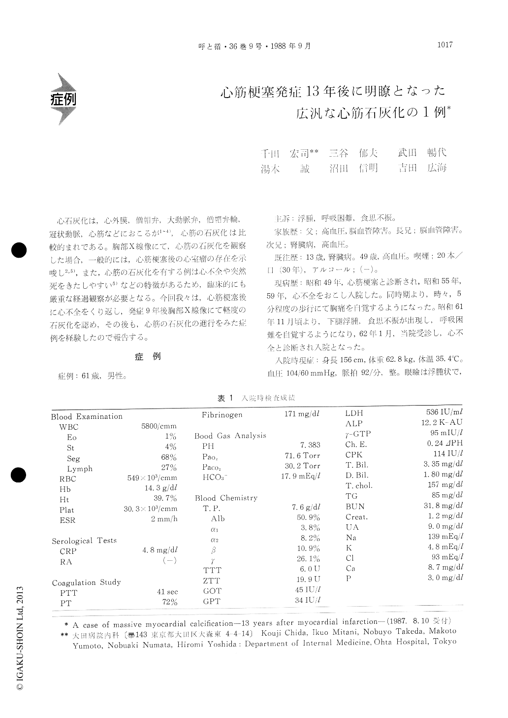
心石灰化は,心外膜,僧帽弁,大動脈弁,僧帽弁輪、冠状動脈心筋などにおこるが1〜4),心筋の石灰化は比較的まれである。胸部X線像にて、心筋の石灰化を観察した場合,一般的には,心筋梗塞後の心室瘤の存在を示唆し2,5),また,心筋の石灰化を有する例は心不全や突然死をきたしやすい5)などの特徴があるため,臨床的にも厳重な経過観察が必要となる。今回我々は,心筋梗塞後に心不全をくり返し,発症9年後胸部X線像にて軽度の石灰化を認め,その後も,心筋の石灰化の進行をみた症例を経験したので報告する。
A 61-year-old man was admitted to this hospital because of edema and dyspnea. There was a history of myocardial infarction (MI) 13 years earlier and two episodes of congestive heart failure seven and three years earlier. Four years before admission X-ray films of the chest revealed a curvelinear and moderate calcification of the left ventricular an-terior wall (AW) on the lateral view, the length of which was about four centimeter. During two mon-ths before admission edema and dyspnea developed. There was++ pretibial edema, and bulbar conjuncti-va was icteric. Diminished breath sounds and wheez-ing were heard at both lungs and coarse crackles at the base of both lungs. The heart was enlarged, with pan-systolic murmur at the apex. ECG revealed the presence of a previous anterior and lateral MI. X-ray films of the chest revealed a curvelinear and massive calcification of the interventricular septum (IVS) and AW including the apex on the lateral and left anterior view, the size of which was 8 × 8 centimeter. M-mode and 2-dimentional echocardio-grams showed a strong echo density of IVS and AW including the apex and the left ventricular posterior wall could not be recorded because of the acoustic shadow due to the stronger echo density of IVS at the level of papillary muscles. Also, computed tomo-graphy of the heart showed the massive myocardial calcification at same sites. There was no evidence of ventricular aneurysm with a protrusion outside cardiac borders.
In this case, it was showed that the myocardial calcification became clear and progressed in com-parison with that before four years.

Copyright © 1988, Igaku-Shoin Ltd. All rights reserved.


