Japanese
English
- 有料閲覧
- Abstract 文献概要
- 1ページ目 Look Inside
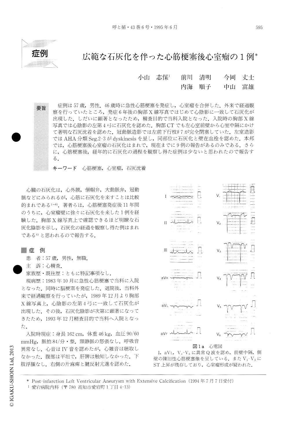
症例は57歳,男性.46歳時に急性心筋梗塞を発症し,心室瘤を合併した.外来で経過観察を行っていたところ,発症6年後の胸部X線写真ではじめて心陰影に一致して石灰化が出現した.しだいに顕著となったため,精査目的で当科入院となった.入院時の胸部X線写真では心陰影の左第4弓に石灰化を認めた.胸部CTでも左心室前壁から心室中隔にかけて著明な石灰沈着を認めた.冠動脈造影では左前下行枝#7が完全閉塞していた.左室造影ではAHA分類Seg.2-3がdyskinesisを呈し,同部位に石灰化と壁在血栓を認めた.本邦では,心筋梗塞後心室瘤の石灰化はまれで,現在までに9例の報告があるのみである.さらに,心筋梗塞後,経年的に石灰化の過程を観察し得た症例は少ないと思われたので報告する.
We reported a case of calcified left ventricular aneur-ysm 11 year after initial myocardial infarction. A 57-year-old man was admitted to our hospital. On chest X-ray and chest CT scan, marked calcification was found in the left ventricle. Calcium deposit was found on chest X-ray six years after the initial infarction. Coronary angiography showed complete obstruction of the left anterior branch seg. 7. Left ventriculography showed abnormal contractility around the center of the apex region.

Copyright © 1995, Igaku-Shoin Ltd. All rights reserved.


