Japanese
English
- 有料閲覧
- Abstract 文献概要
- 1ページ目 Look Inside
心筋梗塞後の心室瘤の頻度は,その定義の差の為報告者によりまちまちである。しかし,一般に心室瘤群は,非心室瘤群に比してその予後は悪いとされている。大動脈内バルーンパンピング法の導入などにより,重症心不全状態も切りぬけられ,長期経過中心室瘤を形成する例が今後増加すると考えられ,臨床的にも梗塞後心室瘤を有する患者の取扱いは重要となっている。また,症例によっては外科的処置も可能であり,その早期診断も重要である1,2)。
今回我々は,広範囲梗塞後心室瘤に著明な石灰化を伴ない14年間生存した1剖検例を経験したので,若干の文献的考察を加えながら検討してみたい。
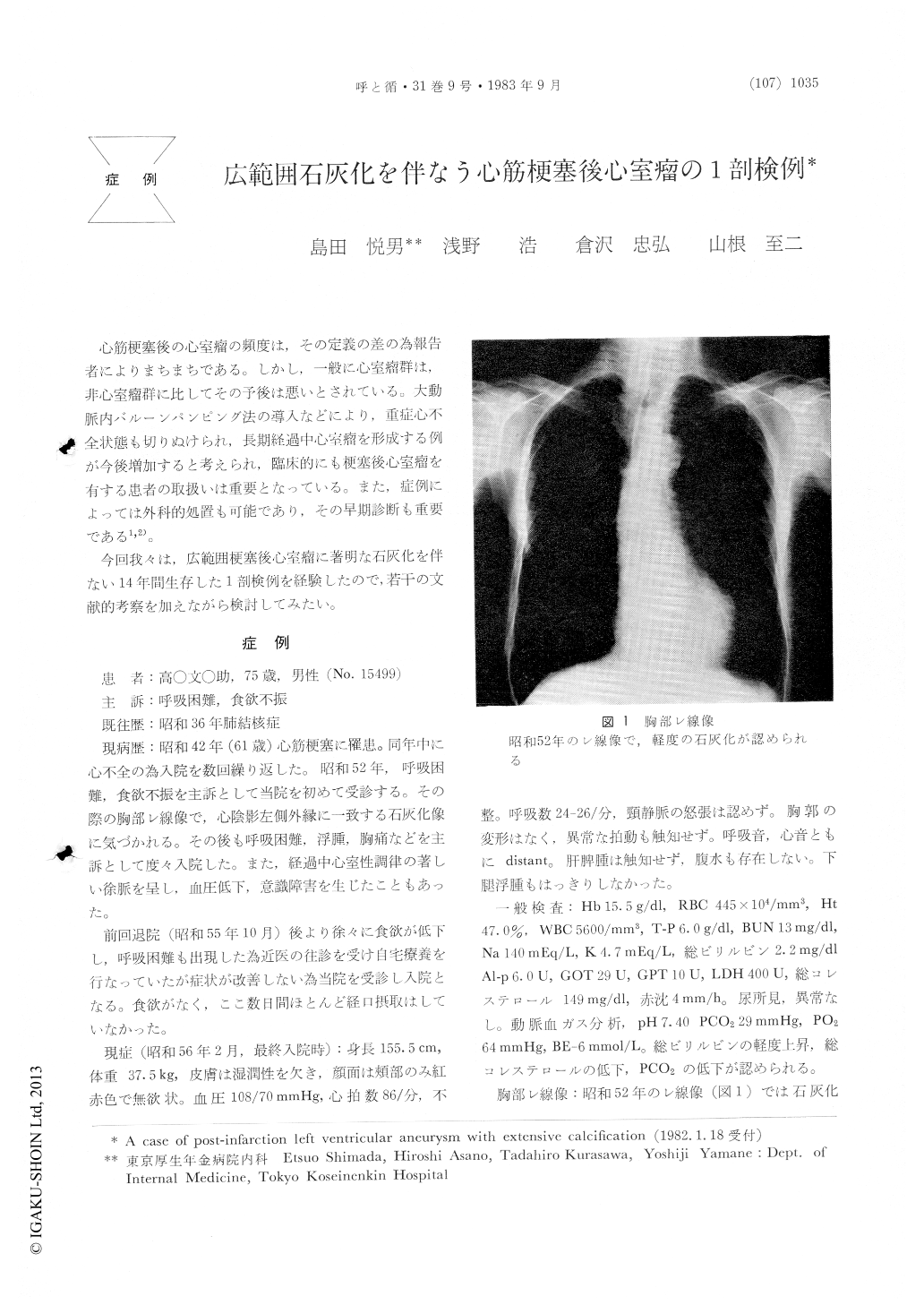
The patient, 75 years old man, suffered an attack of myocardial infarction 14 years previously. In 1977, he was initially admitted on complaint of dyspnea. At that time, the chest x-rays disclosed that the cardiac silhouette was normal in size, and a hemispheric shell of calcific material formed the apex of the left and the inferior margins of the cardiac shadow. On computed tomography, this calcific material confirmed the calcification of the aneurysmal bulge at the apex.
Pathological findings. The heart weighed 340g, the pericardium was firm and fibrous adhesions over the cardiac apex. At the apex of the left ventricle, the myocardium was diminished with the formations of a fibrous aneurysmal bulge. The wall of the aneurysm was hard and rigid due to extensive calcification, and the sac was partially filled by adherent organized thrombus.
A presentation has been made of a case of calcific left ventricular aneurysm in man, aged 75, with an exceptionally long history with cardiac infarction ocurring 14 years previously. A search for published reports of similar cases is uncommon.

Copyright © 1983, Igaku-Shoin Ltd. All rights reserved.


