Japanese
English
- 有料閲覧
- Abstract 文献概要
- 1ページ目 Look Inside
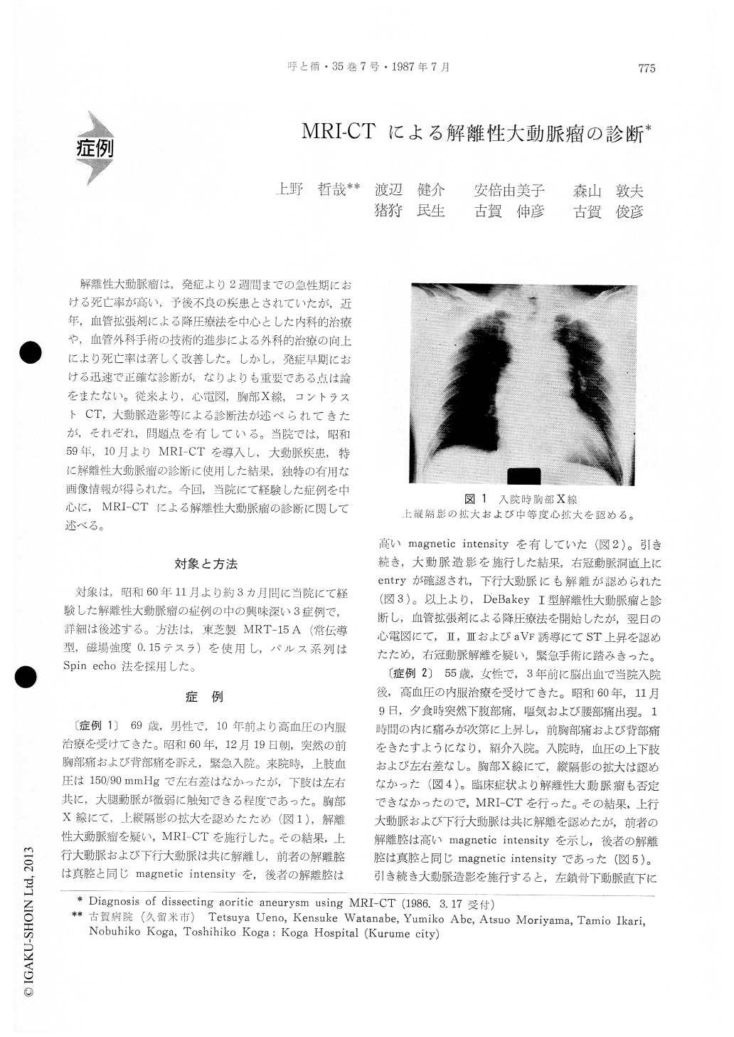
解離性大動脈瘤は,発症より2週間までの急性期における死亡率が高い,予後不良の疾患とされていたが,近年,血管拡張剤による降圧療法を中心とした内科的治療や,血管外科手術の技術的進歩による外科的治療の向上により死亡率は著しく改善した。しかし,発症早期における迅速で正確な診断が,なりよりも重要である点は論をまたない。従来より,心電図,胸部X線,コントラストCT,大動脈造影等による診断法が述べられてきたが,それぞれ,問題点を有している,当院では,昭和59年,10月よりMRI-CTを導入し,大動脈疾患,特に解離性大動脈瘤の診断に使用した結果,独特の有用な画像情報が得られた。今回,当院にて経験した症例を中心に,MRI-CTによる解離性大動脈瘤の診断に関して述べる。
We evaluated the diagnostic efficacy of Nuclear Magnetic Resonance Imaging-Computed Tomography (MRI-CT) for dissecting aortic aneurysm. Through our clinical evaluations using MRI-CT, the following conclusions were obtained.
1) Since the MRI-CT produces clear contrast between the vessel wall and flowing blood that the pathological changes of aortic wall, aortic dissection, and/or aortic aneurysm can be definitely evaluated.
2) The MRI-CT is useful as a noninvasive method for screening of dissecting aortic aneurysm among several diseases which cause severe back pain.
3) The MRI-CT can definitely clarify the distinc-tion of De Bakey type I or type Ill of retrograde aneurysm concerning to the dissecting aorta.
4) For further clinical applications, we need to develop following problems.
a) Faster and finer imaging should be developed.
b Because of the problem of magnetic affection from metalic substances, the technical development is required to check the patients' vital signs during its use.

Copyright © 1987, Igaku-Shoin Ltd. All rights reserved.


