Japanese
English
- 有料閲覧
- Abstract 文献概要
- 1ページ目 Look Inside
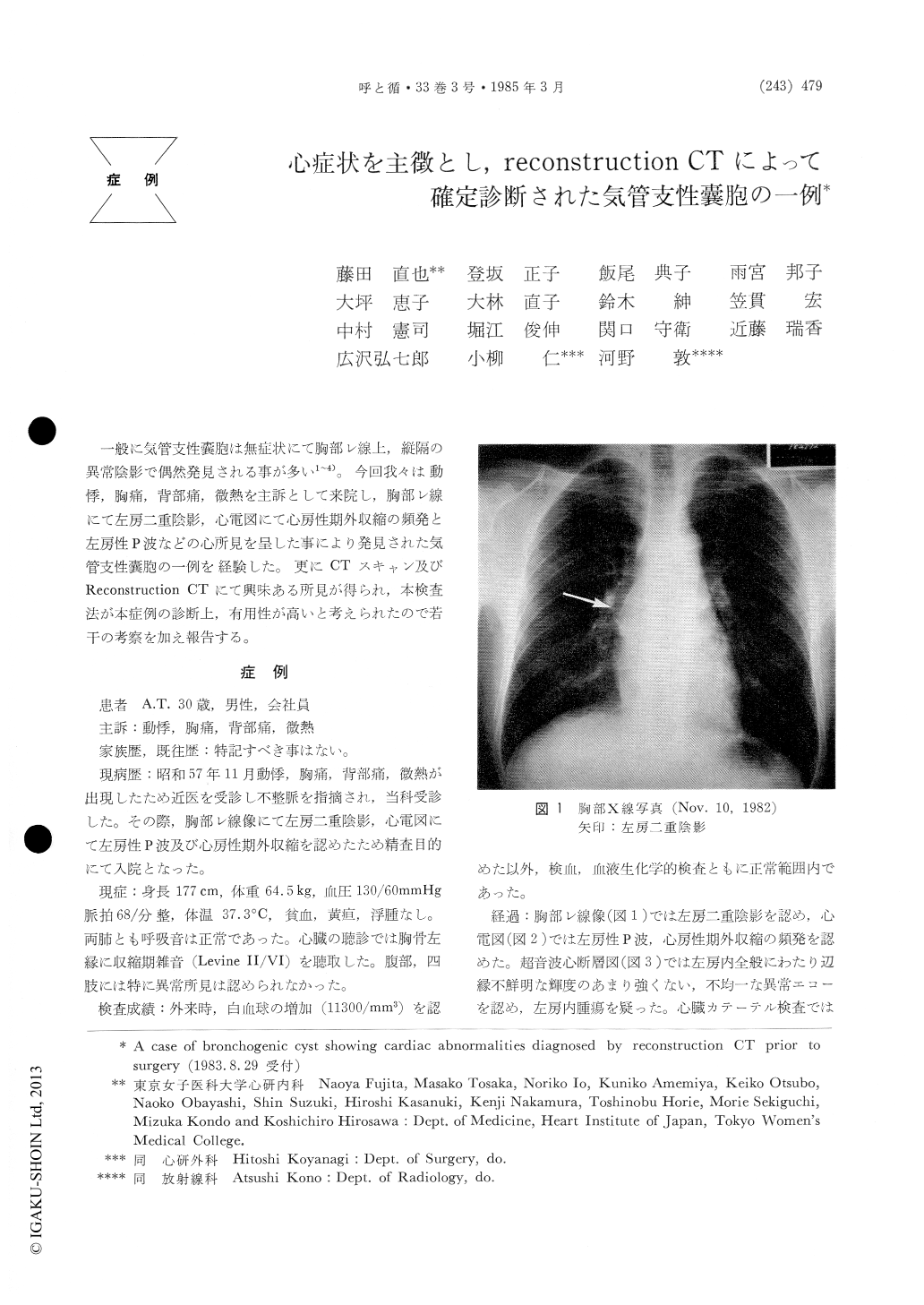
一般に気管支性嚢胞は無症状にて胸部レ線上,縦隔の異常陰影で偶然発見される事が多い1〜4)。今回我々は動悸,胸痛,背部痛,微熱を主訴として来院し,胸部レ線にて左房二重陰影,心電図にて心房性期外収縮の頻発と左房性P波などの心所見を呈した事により発見された気管支性嚢胞の一例を経験した。更にCTスキャン及びReconstruction CTにて興味ある所見が得られ,本検査法が本症例の診断上,有用性が高いと考えられたので若干の考察を加え報告する。
A 30-year-old man began to experience palpita-tion, chest pain and fever 3 weeks prior to admission. An electrocardiogram showed evidence suggestive of left atrial overloading, and frequent atrial premature beats. A chest roentgenogram revealed an abnormal shadow behind the right upper portion of the heart. A cross-sectional echocardiogram demonstrated abnormal echoes with ill-defined margins in the left atrium and a picture of Computed Tomography (CT) was thought to show a left atrial tumor. How-ever left atriography suggested it to be an extra-cardiac tumor which was compressing the left atrium. Therefore, reconstruction CT was perform-ed and with the use of high CT numbers, the cyst was considered to be subcarinal in position. The CT also showed a calcified mass which was mobile within the cyst.
At thoracotomy, an 6×3 cm cyst was removed completely and a diagnosis of bronchogenic cyst was established histopathologically.
In this report, ECG abnormalities suggestive of left atrial damage and the diagnostic usefulness of reconstruction CT are stressed.

Copyright © 1985, Igaku-Shoin Ltd. All rights reserved.


