Japanese
English
研究
左室内血栓検出に関する超音波断層法とCT scanの比較検討
A comparative evaluation of cross-sectional echocardiography and computed tomography in a detection of left ventricular thrombus
金光 弘
1
,
石川 恭三
1
Hiroshi Kanemitsu
1
,
Kyozo Ishikawa
1
1杏林大学第2内科
1Kyorin University school of medicine 2nd department of internal medicine
pp.835-842
発行日 1982年8月15日
Published Date 1982/8/15
DOI https://doi.org/10.11477/mf.1404204065
- 有料閲覧
- Abstract 文献概要
- 1ページ目 Look Inside
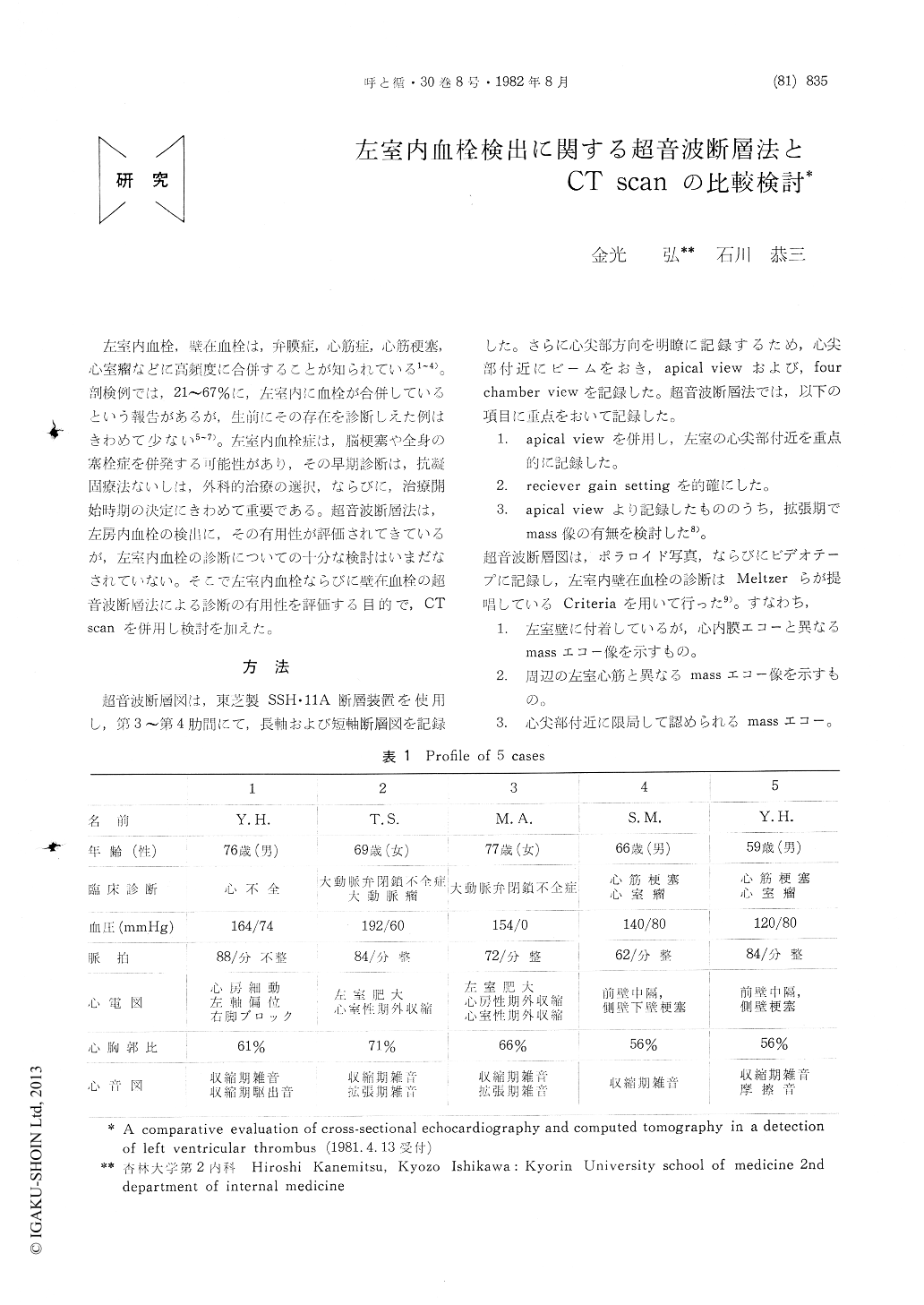
左室内血栓,壁在血栓は,弁膜症,心筋症,心筋梗塞,心室瘤などに高頻度に合併することが知られている1〜4)。剖検例では,21〜67%に,左室内に血栓が合併しているという報告があるが,生前にその存在を診断しえた例はきわめて少ない5〜7)。左室内血栓症は,脳梗塞や全身の塞栓症を併発する可能性があり,その早期診断は,抗凝固療法ないしは,外科的治療の選択,ならびに,治療開始時期の決定にきわめて重要である。超音波断層法は,左房内血栓の検出に,その有用性が評価されてきているが,左室内血栓の診断についての十分な検討はいまだなされていない。そこで左室内血栓ならびに壁在血栓の超音波断層法による診断の有用性を評価する目的で,CT scanを併用し検討を加えた。
Left ventricular (LV) thrombi are rarely re-cognized during life, though they are not in-frequently found at post-mortem examination of patients succumbed to valvular disease, acute myocardial infarction, and cardiomyopathy. We presented five cases in which LV thrombi were detected by cross-sectional echocardiography (CSE) and confirmed by computed tomography.

Copyright © 1982, Igaku-Shoin Ltd. All rights reserved.


