Japanese
English
- 有料閲覧
- Abstract 文献概要
- 1ページ目 Look Inside
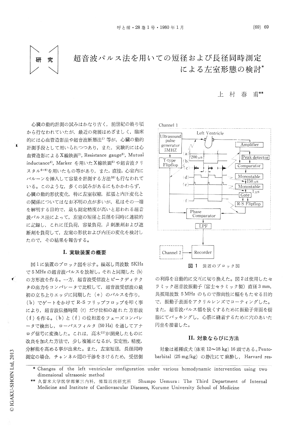
心臓の動的計測の試みはかなり古く,前世紀の終り頃から行なわれていたが,最近の発展はめざましく,臨床的には心血管造影法や超音波断層法1)等が,心臓の動的計測手段として用いられつつあり,また,実験的には心血管造影によるX線映画2),Resistance gauge3),Mutual inductance4),Markerを用いたX線映画4)や超音波クリスタル6〜9)を用いたもの等があり,また,直接,心室内にバルーンを挿入して容量を計測する方法10)も行なわれている。このような,多くの試みがあるにもかかわらず,心臓の動的形状変化,特に左室収縮,拡張と内圧変化との関係についてはなお不明の点が多いが,私はその一端を解明する目的で,最も測定精度が高いと思われる超音波パルス法によって,左室の短径と長径を同時に連続的に記録し,これに圧負荷,容量負荷,β刺激剤および遮断剤を負荷して,左室の形状および内圧の変化を検討したので,その結果を報告する。
Minor and major axis diameters of the left ventricle in the anesthetized open-chest dogs were measured using two paires of pulse-transit ultrasonic dimension transducers.
1) In the control period, the ratio of iso-volumic sphericalization pattern, that is length-ning of minor axis diameter and shorthning of major axis diameter during isovolumic contrac-tion, to isovolumic elipticalization pattern, that is shortning of minor axis diameter and length-ning of major axis diameter during isovolumic contraction, was 1 to 2.

Copyright © 1980, Igaku-Shoin Ltd. All rights reserved.


