Japanese
English
- 有料閲覧
- Abstract 文献概要
- 1ページ目 Look Inside
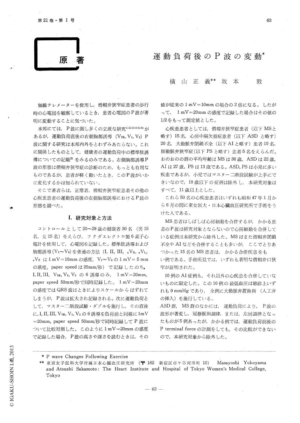
無線テレメーターを使用し,僧帽弁狭窄症患者の歩行時の心電図を観察しているとき,患者心電図のP波が著明に変動することに気づいた。
本邦にては,P波に関し多くの立派な研究1)2)3)4)5)6)があるが,運動負荷直後の右側胸部誘導(V3R, V1, V2) P波に関する研究は本邦内外をとわずみあたらない。これに関係したものとして,健康者の運動負荷中の標準肢誘導についての記載2)をみるのみである。右側胸部誘導P波の形態は僧帽弁狭窄症の診断のため,もっとも有用なものであるが,患者が軽く動いたとき,このP波がいかに変化するかは知られていない。
The P terminal force in V1 in the healthy young is positive both at rest and after exercise.The P terminal force in V1 in patients with mi-tral stenosis was 0.090mm. sec. at rest in the average of 15 cases. The terminal force markedly increased to 0.156mm. sec. following Master single two setp test.
The patients with aortic insufficiency didn't show such prominent changes of the P terminal force as seen in patients with mitral stenosis.In the aortic insufficiency group, the terminal force in V1 was 0.042mm. sec. at rest and 0.050 mm. sec. following the exercise.

Copyright © 1973, Igaku-Shoin Ltd. All rights reserved.


