Japanese
English
- 有料閲覧
- Abstract 文献概要
- 1ページ目 Look Inside
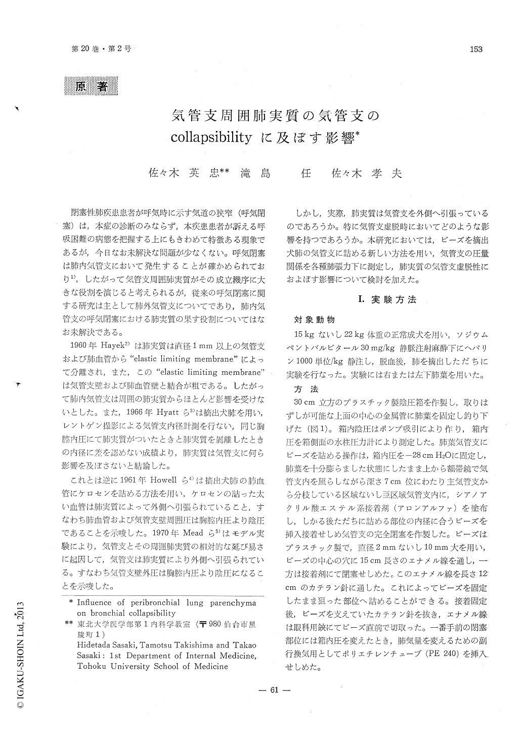
閉塞性肺疾患患者が呼気時に示す気道の狭窄(呼気閉塞)は,本症の診断のみならず,本疾患患者が訴える呼吸困難の病態を把握する上にもきわめて特徴ある現象であるが,今日なお未解決な問題が少なくない。呼気閉塞は肺内気管支において発生することが確かめられており1),したがって気管支周囲肺実質がその成立機序に大きな役割を演じると考えられるが,従来の呼気閉塞に関する研究は主として肺外気管支についてであり,肺内気管支の呼気閉塞における肺実質の果す役割についてはなお未解決である。
1960年Hayek2)は肺実質は直径1mm以上の気管支および肺血管から"elastic limiting membrane"によって分離され,また,この"elastic limiting membrane"は気管支壁および肺血管壁と結合が粗である。したがって肺内気管支は周囲の肺実質からほとんど影響を受けないとした。また,1966年Hyattら3)は摘出犬肺を用い,レントゲン撮影による気管支内径計測を行ない,同じ胸腔内圧にて肺実質がついたときと肺実質を刺離したときの内径に差を認めない成績より,肺実質は気管支に何ら影響を及ぼさないと結論した。
To measure mechanical influence of lung par-enchyma on bronchial collapsiblity, with the ex-cised dog lobe distended at 28 cm H2O of trans-pulmonary pressure, several small beads were in-serted into the bronchi roughly at their 3rd branches and cemented airtight to the bronchial wall with adhesive substance, except one opened to atmosphere with a catheter to obtain different lung expansion through collateral channels with-in the lung.

Copyright © 1972, Igaku-Shoin Ltd. All rights reserved.


