Japanese
English
- 有料閲覧
- Abstract 文献概要
- 1ページ目 Look Inside
- 参考文献 Reference
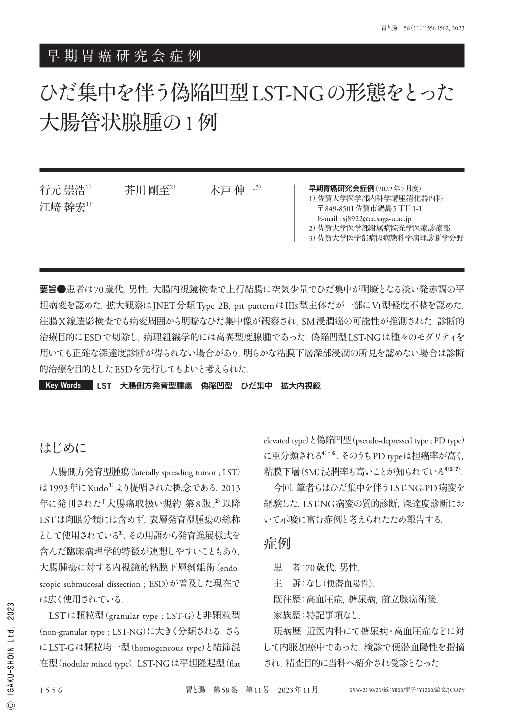
要旨●患者は70歳代,男性.大腸内視鏡検査で上行結腸に空気少量でひだ集中が明瞭となる淡い発赤調の平坦病変を認めた.拡大観察はJNET分類Type 2B,pit patternはIIIS型主体だが一部にVI型軽度不整を認めた.注腸X線造影検査でも病変周囲から明瞭なひだ集中像が観察され,SM浸潤癌の可能性が推測された.診断的治療目的にESDで切除し,病理組織学的には高異型度腺腫であった.偽陥凹型LST-NGは種々のモダリティを用いても正確な深達度診断が得られない場合があり,明らかな粘膜下層深部浸潤の所見を認めない場合は診断的治療を目的としたESDを先行してもよいと考えられた.
An old man in his seventies was detected with a slightly reddish flat elevation with fold convergency in his ascending colon by ileocolonoscopy. Under magnifying colonoscopy using NBI(narrow-band imaging)observation, the microvessel pattern of the tumor was classified as type 2B as per The Japan NBI Expert Team classification. The magnifying observation using crystal violet staining identified the majority of the tumor pit pattern as type III, whereas type VI pit pattern was concurrently detected in some parts. A barium enema examination depicted the lesion as flat depression with vague barium flecks with apparent fold convergency, suggesting a submucosal invasion of the tumor. Histopathological examination of the endoscopically resected specimen revealed that the tumor was tubular adenoma of high-grade atypia with apparent inflammatory cells infiltration in the submucosa. Thus, submucosal invasion was not observed. Considering the difficulty in the diagnosis of preoperative invasion depth of the pseudo-depressed type of LST-NG, ESD can be a choice of treatment when the tumor is suspected as early colorectal cancer.

Copyright © 2023, Igaku-Shoin Ltd. All rights reserved.


