Japanese
English
- 有料閲覧
PPV購入案内
1,320円 (1,200円+税10%) こちらは、1885ページから1890ページの文献です。購入には医書.jp本会員登録が必要です。
会員登録完了後に改めて上記「購入サイトへ」を選択してください。
または「購入サイトへ」選択後に「購入ログイン」、「新規会員登録」の順に進んでください。
会員登録について詳しくはをご確認ください。
- Abstract 文献概要
- 1ページ目 Look Inside
- 参考文献 Reference
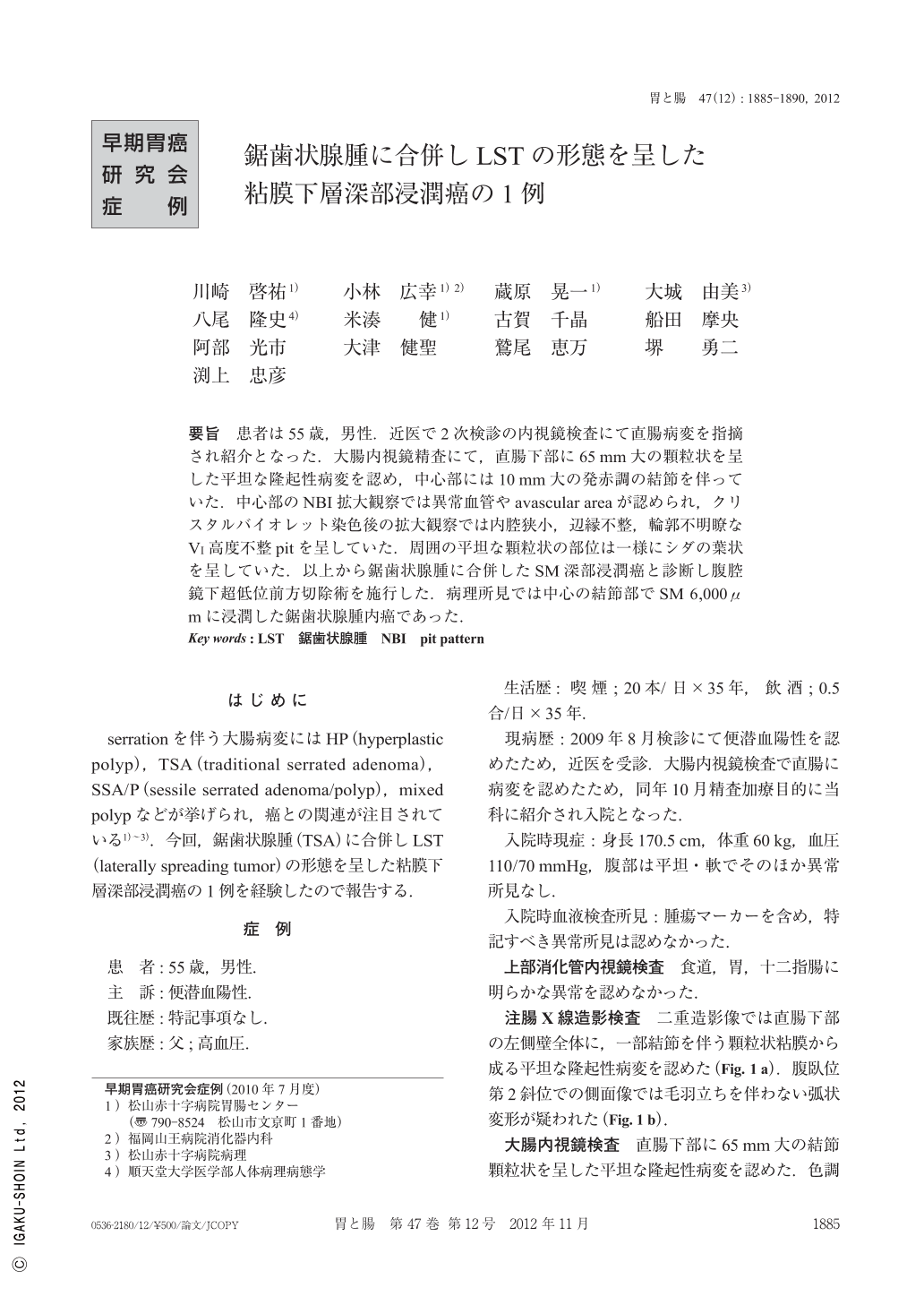
要旨 患者は55歳,男性.近医で2次検診の内視鏡検査にて直腸病変を指摘され紹介となった.大腸内視鏡精査にて,直腸下部に65mm大の顆粒状を呈した平坦な隆起性病変を認め,中心部には10mm大の発赤調の結節を伴っていた.中心部のNBI拡大観察では異常血管やavascular areaが認められ,クリスタルバイオレット染色後の拡大観察では内腔狭小,辺縁不整,輪郭不明瞭なVi高度不整pitを呈していた.周囲の平坦な顆粒状の部位は一様にシダの葉状を呈していた.以上から鋸歯状腺腫に合併したSM深部浸潤癌と診断し腹腔鏡下超低位前方切除術を施行した.病理所見では中心の結節部でSM 6,000μmに浸潤した鋸歯状腺腫内癌であった.
A 55-year-old man was admitted to our institution because of a rectal lesion detected by colonoscopy carried out on further examination for positive fecal occult blood.
Colonoscopy disclosed a flat elevated lesion with one large nodule, whose surface was granular, measuring 65mm in size, in the lower rectum. The size of the large nodule was 10mm, and was reddish in color. NBI(narrow band imaging)view showed irregular microvessel features and an avascular area in the large nodule. Magnifying endoscopy with crystal violet staining demonstrated highly irregular Vi pit pattern within the large nodule. Fern-like patterns were seen in the surrounding flat, granular area. The tentative endoscopic diagnosis of the lesion was submucosal invasive cancer. The lesion was resected surgically by super low anterior resection of the rectum. Histological examination of the resected specimen showed well-to-moderately differentiated tubular adenocarcinoma in a serrated adenoma. Depth of invasion was 6,000μm in the large nodule. We diagnosed this lesion to be an early colorectal cancer arising in a serrated adenoma.
Copyright © 2012, Igaku-Shoin Ltd. All rights reserved.