Japanese
English
- 有料閲覧
- Abstract 文献概要
- 1ページ目 Look Inside
- 参考文献 Reference
- サイト内被引用 Cited by
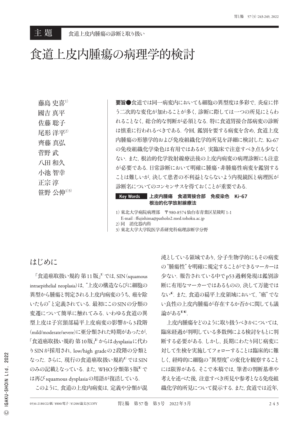
要旨●食道では同一病変内においても細胞の異型度は多彩で,炎症に伴う二次的な変化が加わることが多く,診断に際しては一つの所見にとらわれることなく,総合的な判断が必須となる.特に食道胃接合部病変の診断は慎重に行われるべきである.今回,鑑別を要する病変を含め,食道上皮内腫瘍の形態学的および免疫組織化学的所見を詳細に検討した.Ki-67の免疫組織化学染色は有用ではあるが,実臨床で注意すべき点も少なくない.また,根治的化学放射線療法後の上皮内病変の病理診断にも注意が必要である.日常診断において明確に腫瘍・非腫瘍性病変を鑑別することは難しいが,決して患者の不利益とならないよう内視鏡医と病理医が診断名についてのコンセンサスを得ておくことが重要である.
The esophagus contains various atypia even within the same lesion, and many secondary changes occur because of inflammation. Therefore, diagnosis should be based on a comprehensive evaluation and not on a single finding. We reviewed the morphological and immunohistological properties of lesions that differentiate them from tumors. Although Ki-67 is a useful marker, many cautions must be taken. In recent years, many follow-up cases after definitive chemoradiation have been reported, and caution is required. Although tumors and nontumors are difficult to differentiate clearly in routine diagnosis, endoscopists and pathologists must reach a consensus on the diagnostic term so that patients are not disadvantaged.

Copyright © 2022, Igaku-Shoin Ltd. All rights reserved.


