Japanese
English
- 有料閲覧
- Abstract 文献概要
- 1ページ目 Look Inside
- 参考文献 Reference
- サイト内被引用 Cited by
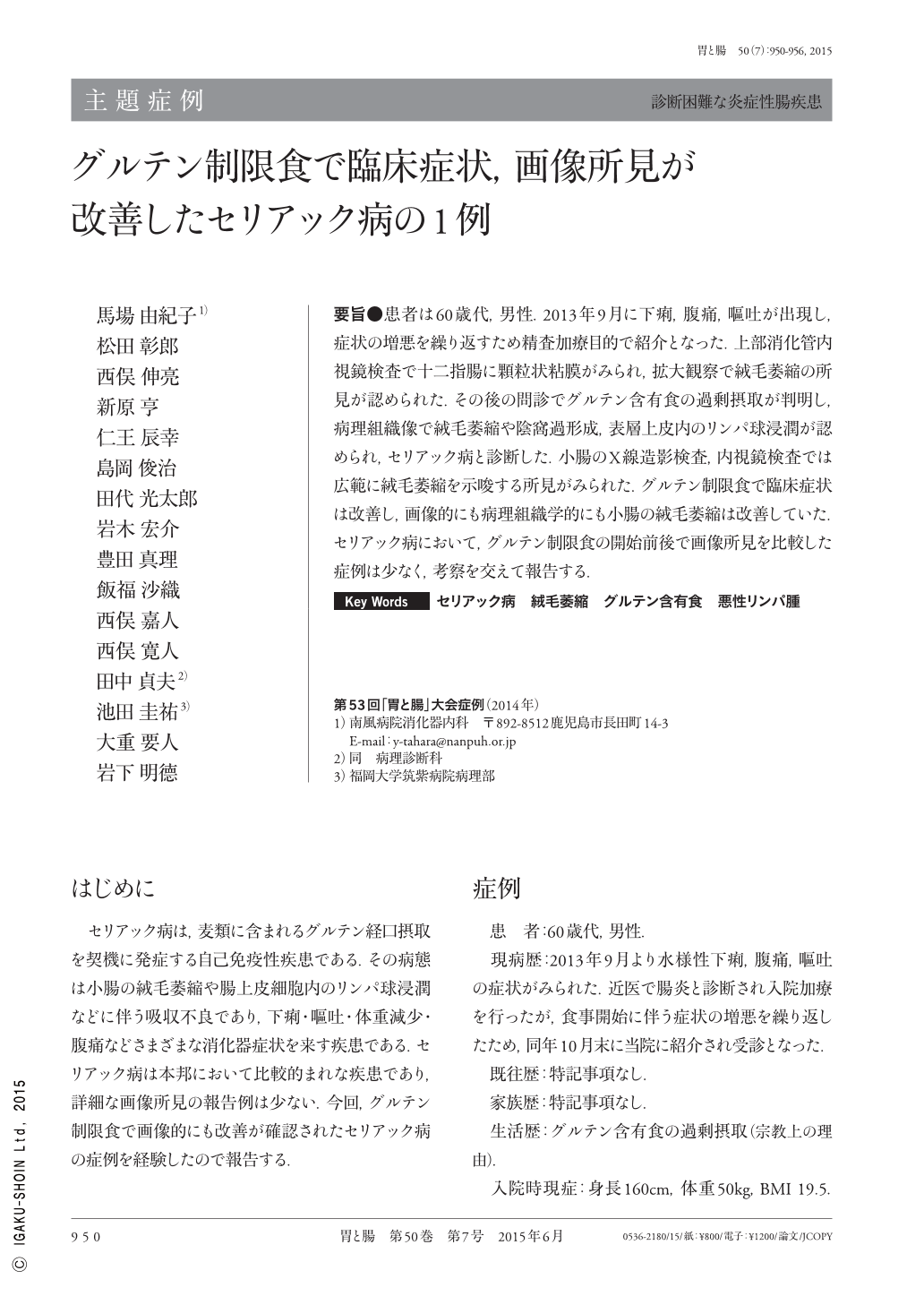
要旨●患者は60歳代,男性.2013年9月に下痢,腹痛,嘔吐が出現し,症状の増悪を繰り返すため精査加療目的で紹介となった.上部消化管内視鏡検査で十二指腸に顆粒状粘膜がみられ,拡大観察で絨毛萎縮の所見が認められた.その後の問診でグルテン含有食の過剰摂取が判明し,病理組織像で絨毛萎縮や陰窩過形成,表層上皮内のリンパ球浸潤が認められ,セリアック病と診断した.小腸のX線造影検査,内視鏡検査では広範に絨毛萎縮を示唆する所見がみられた.グルテン制限食で臨床症状は改善し,画像的にも病理組織学的にも小腸の絨毛萎縮は改善していた.セリアック病において,グルテン制限食の開始前後で画像所見を比較した症例は少なく,考察を交えて報告する.
In September 2013, a male patient in his 60's experienced diarrhea, abdominal pain, and vomiting, which repeatedly exacerbated. Therefore, he was referred to our hospital for detailed examination and treatment. Upper gastrointestinal endoscopy revealed granular mucosa in the duodenum. Magnification showed findings of villous atrophy. Following a medical interview, it was understood that the patient consumed an excessive amount of gluten. Histopathological findings indicated villous atrophy, crypt hyperplasia, and lymphocyte infiltration within the surface epithelium, leading to a diagnosis of celiac disease. Small bowel X-ray examination and endoscopy revealed findings suggestive of extensive villous atrophy. A gluten-free diet was prescribed, after which the clinical symptoms improved. Furthermore, imaging and histopathological findings demonstrated that the small bowel villous atrophy improved. There are few reports in the literature that have compared imaging findings before and after the implementation of a gluten-free diet;this report will hereby present this case together with a discussion.

Copyright © 2015, Igaku-Shoin Ltd. All rights reserved.


