Japanese
English
- 有料閲覧
- Abstract 文献概要
- 1ページ目 Look Inside
胃癌の肉眼所見は,その病理組織構築を反映し,X線,内視鏡検査はこの肉眼所見を忠実に描出してその病理組織構築を推定することにある.
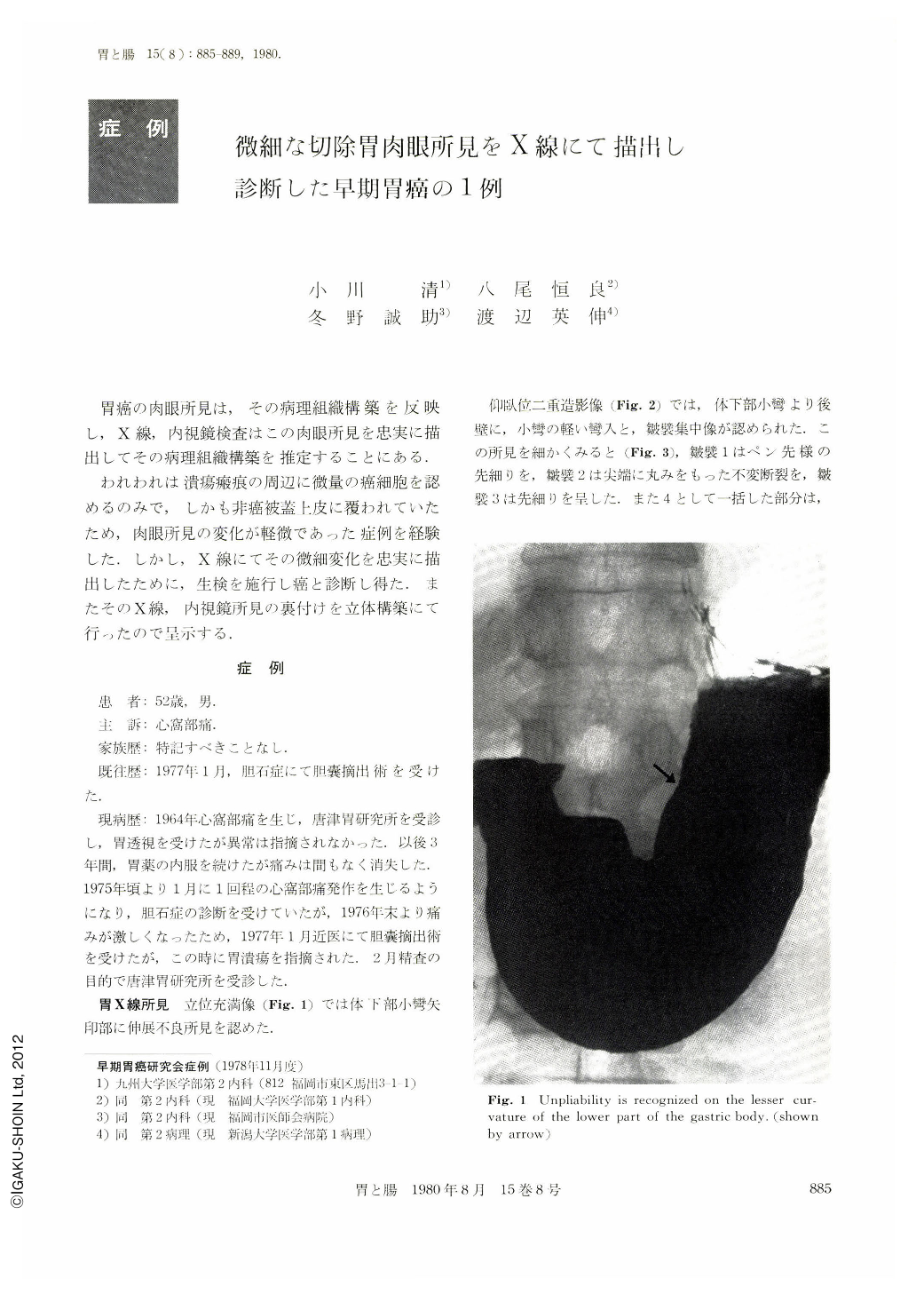
われわれは潰瘍瘢痕の周辺に微量の癌細胞を認めるのみで,しかも非癌被蓋上皮に覆われていたため,肉眼所見の変化が軽微であった症例を経験した.しかし,X線にてその微細変化を忠実に描出したために,生検を施行し癌と診断し得た.またそのX線,内視鏡所見の裏付けを立体構築にて行ったので呈示する.
A 52 year-old man visited Karatsu Cancer Research Institute because of epigastralgia in February 1977. X-ray examination of the stomach disclosed a depressed lesion with folds' convergence on the posterior wall of the lower part of the body and a part of the converging folds was suggestive of malignancy. On endoscopic examination there was no finding suggestive of malignancy, but poorly differentiated adenocarcinoma was confirmed by histological examination of biopsy specimens.
Macroscopic findings of the resected specimen were compatible with those of x-ray films. Microscopically the lesion was almost covered with non-cancerous covering epithelia and a small number of cancer cells were found around an ulcer scar (Ul-IV). It was speculated that most of cancer cells dropped out in the course of the malignant cycle and a few cancer cells were left around the ulcer scar.

Copyright © 1980, Igaku-Shoin Ltd. All rights reserved.


