Japanese
English
- 有料閲覧
- Abstract 文献概要
- 1ページ目 Look Inside
小腸悪性腫瘍は比較的稀であり1),近年の小腸X線診断学の著しい進歩にもかかわらず,その性状診断は必ずしも容易とはいえない.
われわれは最近,下血を主訴とし限局性腫瘤の表面に潰瘍を伴った回腸悪性リンパ腫の1例を経験したので症例を報告し,主として診断学的な立場から若干の考察を加えてみたい.
症 例
患者:27歳,男性,会社員
主訴:下血・下痢
既往歴・生活歴:特記すべきことなし
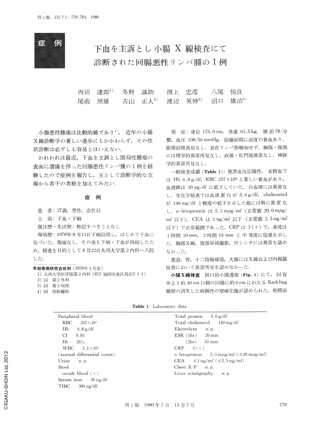
現病歴:1978年9月13日下痢出現し,はじめて下血に気づいた.腹痛なし.その後も下痢・下血が持続したため,精査を目的として9月22日九州大学第2内科へ入院した.
A 24 year-old man was admitted to our clinic because of melena. On admission, both an upper gastointestinal series and a barium enema were negative.
Barium-filled picture of small intestine, however, disclosed abnormalities in the distal portion of the ileum, 6 cm in length and 40 cm oral from the terminal ileum, in which the margin was rigid and Kerkling's folds were abolished. The compression study revealed scant accumulation of the barium surrounded by irregular defects of the shadow, suggesting ulceration in tumor formation. The surface of the tumor appeared slightly uneven, and the displacement of the adjacent loops of small intestine demonstrated on the double contrast film, was indicative of an encircled extraluminal growing of this tumor.
These roentgenological findings strongly suggested malignant lymphoma in the distal ileum. In addition, the superior mesenteric arteriography disclosing smooth encasement of branches of ileal artery by the tumor and faint neovascularies might indicate involvement of this disease in the ileum and also the mesenterium.
On the 30th day of hospitalization, laparotomy was performed and large masses were found in the mesenteric lymph nodes and paraaortic regions besides the tumor of the distal ileum. The tumor was resected and the biopsy of the mesenteric lymph nodes was made.
The resected tumor appeared grossly elastic, 7×4.5 cm in size, and creamy-gray in color. Irregular ulceration was seen on the surface of the tumor. The histological study revealed that the tumor was composed of abundant cellular components. Tumor cells, noted in the submucosal to the muscular layer and infiltrated to the serosa, were large in size having reticular or round to oval nuclei and clear nucleoli. The histological diagnosis was diffuse histiocytic lymphoma.
In this case, the tumor of the distal ileum was thought to be metastic in origin of systemic malignant lymphoma rather than primary localized lymphoma in the ileum.

Copyright © 1980, Igaku-Shoin Ltd. All rights reserved.


