Japanese
English
- 有料閲覧
- Abstract 文献概要
- 1ページ目 Look Inside
- サイト内被引用 Cited by
胃疾患に対する各種診断法の最近の進歩発達はめざましい.1966年第4回日本内視鏡学会秋季大会では2cm以下の胃癌の,1969年第7回日本内視鏡学会秋季大会では1cm以下の微小胃癌の診断が検討され,胃癌に対するX線診断の限界も,肉眼的診断の限界にまで迫らんとする勢いである.また一方では,Ⅱb~Ⅱb類似の表面凹凸の少ない胃癌の診断が,重要な課題としてクローズアップされつつあり,最近では,術前に性状診断されたⅡb病変の報告もみられる.しかしながら,微小胃癌や,Ⅱb~Ⅱb類似病変の,X線内視鏡単独,あるいは両者を併用しても,その性状診断は未だ困難なのが現状で,生検診断の重要性が強調されている.しかしながら,スクリーニング検査の段階で,見落されたり,良性と判定されて,生検診断の網目をくぐりぬけるものがないわけではない.また悪性疑診を置かれていても,微小な病巣のばあい,あるいは病巣と判定された部のごく一部にしか癌が認められない病変では,false negativeを出すことが報告されており,X線・内視鏡による性状診断,癌巣の拡がりの判定に関しては,より一層の精密度が要求されているわけである.したがって,病理組織学的な裏づけを基本にして,X線診断と内視鏡診断をより一層緊密にむすび合わせた,綜合診断学の確立が当面の課題ではなかろうか.
筆者らの経験した早期胃癌症例をふり返えってみると,初期の頃のX線写真は病巣の描出が不十分で,微細な検討に耐えるものは少ない.しかし,次第に小さな,凹凸変化の小さな胃癌が発見されるにつれて,細かい所見が描出されるようになり,最近の症例では,微小な胃癌でも,その微細な変化がよくとらえられており,肉眼標本との対比検討が,十分微細な所までできるようになっている.したがって,肉眼標本に関する微細な所見の検討で得られた知見が,直接X線診断に生かされることが多い.そういった意味から,今回は肉眼標本を対象として検討を行ない,X線診断における,性状診断,微細診断に役立てようと試みた.またX線診断と内視鏡診断をより一層緊密につなぎ合わせる意味で,肉眼標本上の所見と内視鏡上の色調とのむすびつき,さらには癌の組織型との関連についても検討を加えて見た.
Gross specimens of the resected stomachs in 85 cases, 86 lesions, including 66 cases, 67 lesions of depressed type early gastric cancer measuring less than 3cm in the greatest diameter, and 19 cases, 19 lesions of advanced gastric cancer closely resembling early one, have been studied in order to get some clues to diagnostic features of depressed variety of early gastric cancer. The results are as follows:
1) As the borderline of Ⅱc lesion lies next to the areae gastricae half fallen off through cancer nfiltration, irregular surface is produced there (this is called a moth-eaten picture). This finding was uniformly observed.
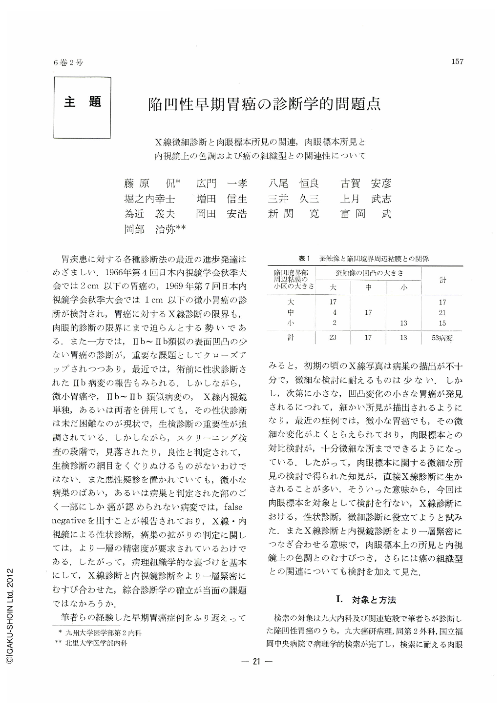
2) Dimensions of unevennes in the moth-eaten area almost coincide with those of the areae gastricae in the surrounding mucosa adjoining the border of depression.
3) Consequently, where the areae gastricae adjacent to depression are small, unevenness at the margin of depression is likewise slight. The marginal line becomes then continuous and smoother.
4) When a mucosal fold is broken off at its tip, the Ⅱa area next to it is fiat, leaving no trace of intact areae gastricae thereon.
5) A cancer lesion seen by endoscopy as an engorgernent mostly belongs to adenocarcinoma. The lesion is rough to smooth with hardly any residue of the areae gastricae.
6) A cancer lesion seen as a discolored area endoscopically is mostly undifferentiated carcinoma. Macroscopically the lesion often shows residues of mucosal folds and areae gastricae.
7) In benign lesions, non-cancerous mucosa seen as an engorged area by endoscopy usually shows regular areae gastricae or a picture of surface microconvergence.

Copyright © 1971, Igaku-Shoin Ltd. All rights reserved.


