Japanese
English
- 有料閲覧
- Abstract 文献概要
- 1ページ目 Look Inside
X線所見,内視鏡所見共に盲腸癌との鑑別が困難であった興味ある回腸末端部脂肪腫の1例を経験したので若干の文献的考察を加えて報告する.
症 例
患 者:花○利○,71歳,男.
主 訴:上腹部痛,右季肋部腫瘤.
既往歴,家族歴:特記すべきことなし.
現病歴:4年ぐらい前から時々上腹部痛があり,そのつど開業医を受診し胃透視を受けたが源因は不明であった.1978年6月ごろより右季肋部に腫瘤を認め,1978年10月25日から再度上腹部痛を来した.とう痛がとれないため3日後に入院した.
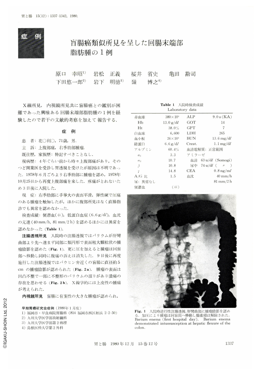
A 71 year-old man was admitted to Sawara Hospital complaining of both intermittent severe epigastric pain and of the tumor palpation at right hypochondrium. Barium enema demonstrated intussusception at hepatic flexure of the colon, and a tumor about 5 cm in diameter was disclosed at the cecum after the reduction. The surface of the tumor was slightly lobulated and irregularly-shaped small ulceration was found on its top. Endoscopically, similar findings were obtained and a stalk was also revealed. Epithelial tumor, especially cancer of the cecum, was suspected because of redness and ulceration of the surface as well as its size but biopsy revealed intact mucosa instead of malignant tissue.
Laparotomy was performed against the repeated attacks of the intussusception under the diagnosis of submucosal tumor. Right hemicolectomy was performed including terminal ileum of about 10 cm towards the oral side from ileocecal valve. Resected specimen showed that the tumor,3.8×3.2×5 cm in size, was derived from the terminal ileum near the valve. Histologically, tumor was comprised of the encapsulated well differentiated adipose tissue and was located at the submucosa. The surface of the tumor was completely covered with ileal mucosa except for the part of ulceration on its top. In differentiating submucosal lipoma from cancer, it should be noted that variegated inflammatory change could occur at the surface after the repeated intussusception or expansion by tumor growth.

Copyright © 1980, Igaku-Shoin Ltd. All rights reserved.


