Japanese
English
- 有料閲覧
- Abstract 文献概要
- 1ページ目 Look Inside
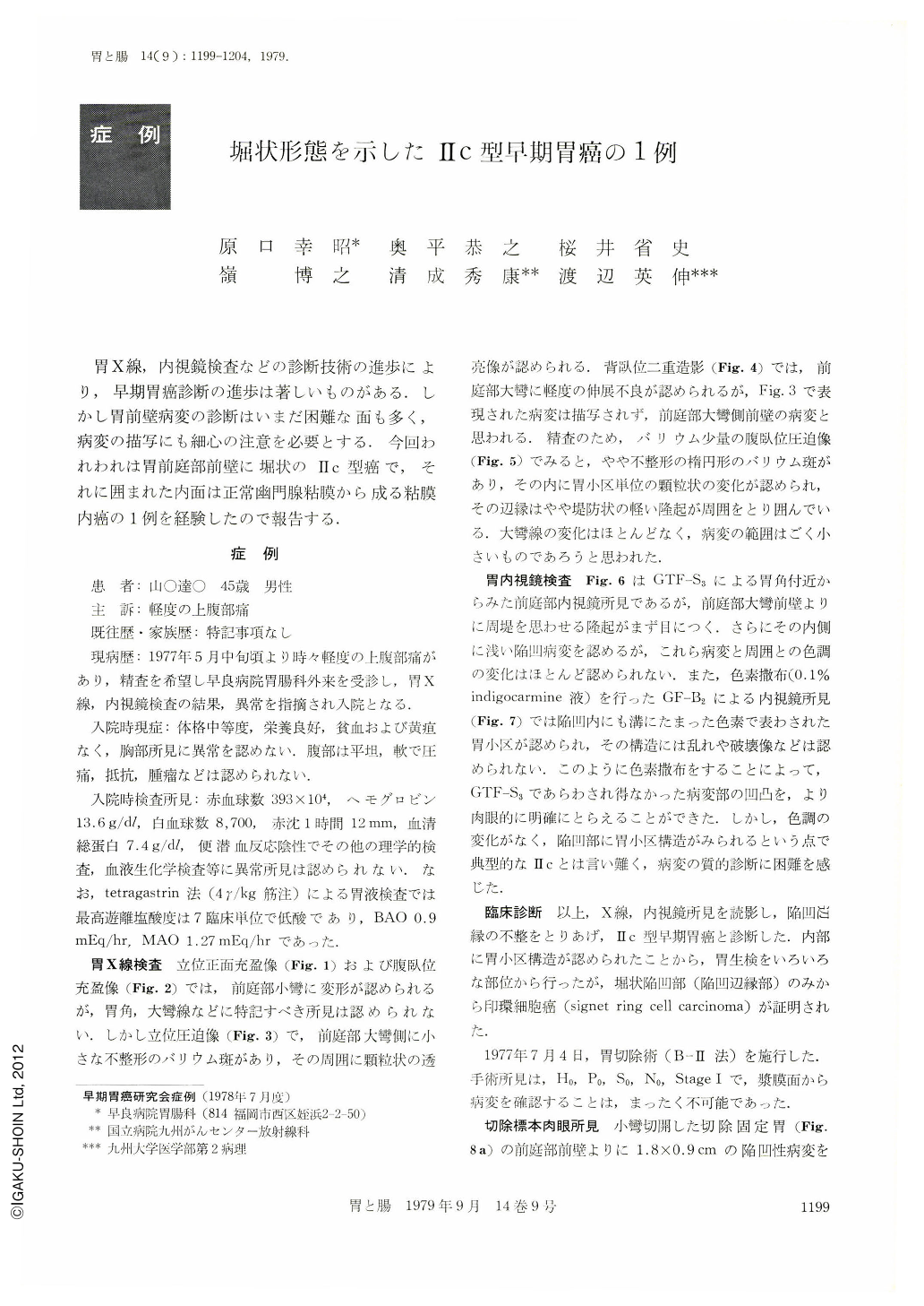
胃X線,内視鏡検査などの診断技術の進歩により,早期胃癌診断の進歩は著しいものがある.しかし胃前壁病変の診断はいまだ困難な面も多く,病変の描写にも細心の注意を必要とする.今回われわれは胃前庭部前壁に堀状のⅡc型癌で,それに囲まれた内面は正常幽門腺粘膜から成る粘膜内癌の1例を経験したので報告する.
Patient: A 45 years old male.
The examination of the stomach revealed a small irregularly-shaped barium fleck with marginal granular elevation on the anterior wall of the antrum. Compression pictures of the lesion in the prone position demonstrated small granular finding similar to normal gastric areas. A gastrofiberscopic examination showed the same lesion as was revealed by the X-ray examination and we distinctly recognized normal gastric areas in that depression by the dyeing technique with indigocarmine solution. There was no difference in color tone between the lesion and the normal gastric mucosa.
The lesion was not typical of 11c type gastric carcinoma. Biopsy specimens picked up from erosions in moat-like narrow depression at the periphery of the lesion revealed signet ring cell carcinoma.
In the resected stomach, a moat-like Ⅱc carcinoma, 1.8×0.9cm, with normal gastric areas in the wide central areas was present in the anterior wall of the antrum. Signet ring cell carcinoma was found only in the narrow moat and limited to the mucosa. The area surrounded by the moat-like Ⅱc carcinoma was composed of almost normal pyloric mucosa. This type of Ⅱc carcinoma is very rare and histologically consists of signet ring cell carcinoma. Clinically, it is of most importance to find a moat-like depression with occasional erosions, which are the most rewarding area for an endoscopic biopsy.

Copyright © 1979, Igaku-Shoin Ltd. All rights reserved.


