Japanese
English
- 有料閲覧
- Abstract 文献概要
- 1ページ目 Look Inside
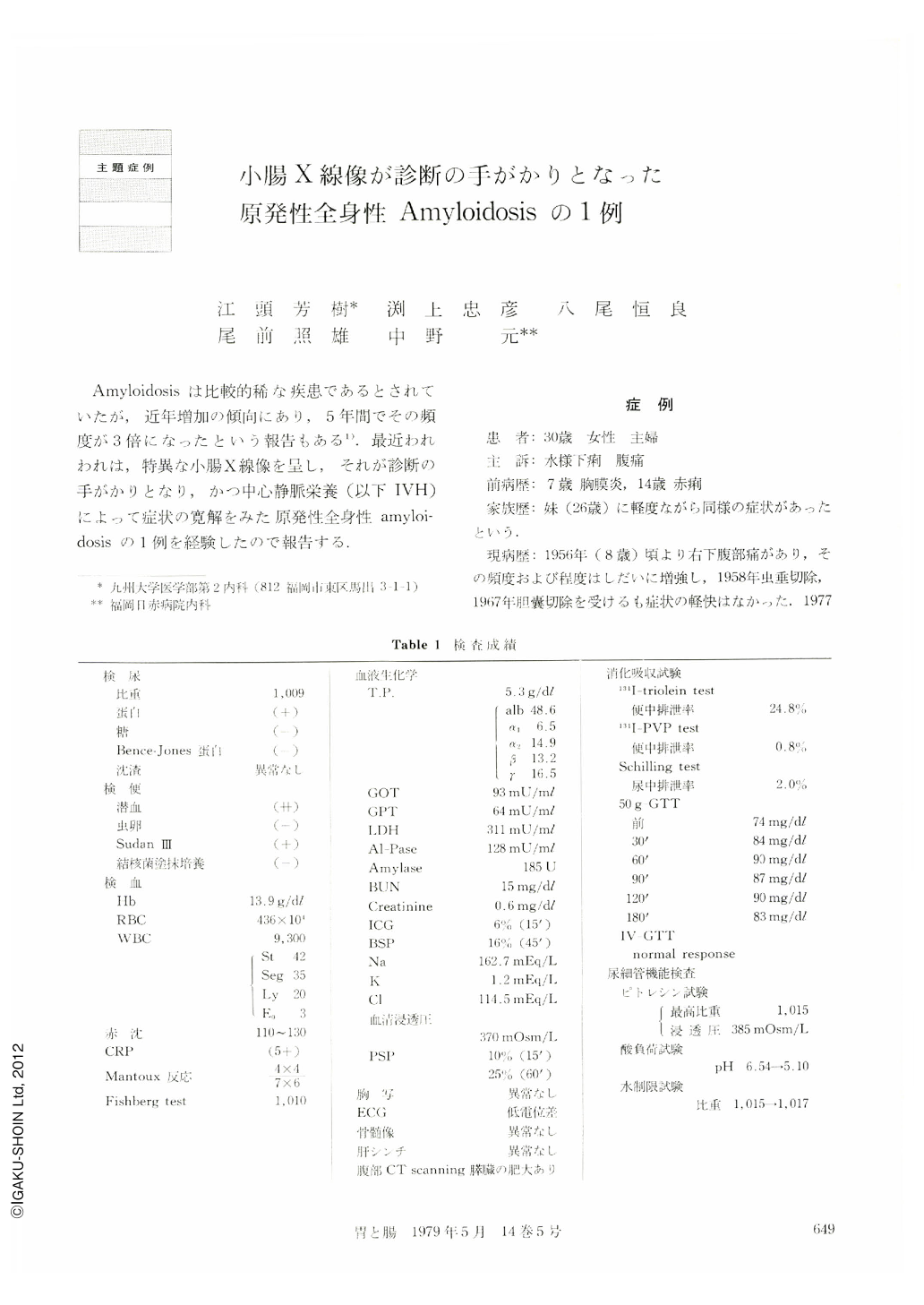
Amyloidosisは比較的稀な疾患であるとされていたが,近年増加の傾向にあり,5年間でその頻度が3倍になったという報告もある1).最近われわれは,特異な小腸X線像を呈し,それが診断の手がかりとなり,かつ中心静脈栄養(以下IVH)によって症状の寛解をみた原発性全身性amyloidosisの1例を経験したので報告する.
A 30-year-old female had been suffering from abdominal pain since 1956. Watery diarrhea developed in 1977 in association with pregnancy and she was admitted to Kyushu University Hospital.
The skin was dry and the turgor was poor. Intestinal loop was palpable and tenderness was felt in the right lower quadrant of the abdomen.
Occult blood of feces was continuously positive; ESR was accelerated and CRP was strongly positive. Remarkable dehydration, hypoproteinemia and disturbance of absorption were recognized. Radiography of the small intestine revealed prolonged transit time, decrease of the tonus and disturbed transformability of the mucosa. Edematous thickening of the folds, coarse mucosal appearance, spotty barium flecks and granular elevations were also recognized. Above all, disturbed transformability of the mucosa made us think of amyloidosis and the diagnosis was confirmed by rectal biopsy. Amyloid deposition was also proved in the biopsy specimens of the stomach, duodenum, jejunum and kidney. The ileum, heart, liver and pancreas were suspected to be involved.
Hyperalimentation was performed. Thereafter subjective symptoms lessened and occult blood of feces and CRP became negative. It is speculated that the improvement was caused by recanalization of the vessels in the submucosa and mucosal regeneration induced by the rest of the intestine during hyperalimentation. Radiographic findings of the small intestine revealed remarkable improvement; however, disturbed transformability of the mucosa and coarse mucosal appearance still remained. It is considered that the findings caused by hypoproteinemia and intestinal inflammation were improved and that only those caused by amyloid deposition were left. Therefore, we think that the radiographic appearance of intestinal amyloidosis written in the literature may be influenced by miscellaneous factors. If fine changes can be interpreted in the double contrast study of the small intestine, diagnosis of intestinal amyloidosis may be made as in the diseases of the stomach and colon from the shape of ulcer and fine mucosal appearance not only gross appearance such as disturbed transformability of the mucosa.
Copyright © 1979, Igaku-Shoin Ltd. All rights reserved.