Japanese
English
- 有料閲覧
- Abstract 文献概要
- 1ページ目 Look Inside
- サイト内被引用 Cited by
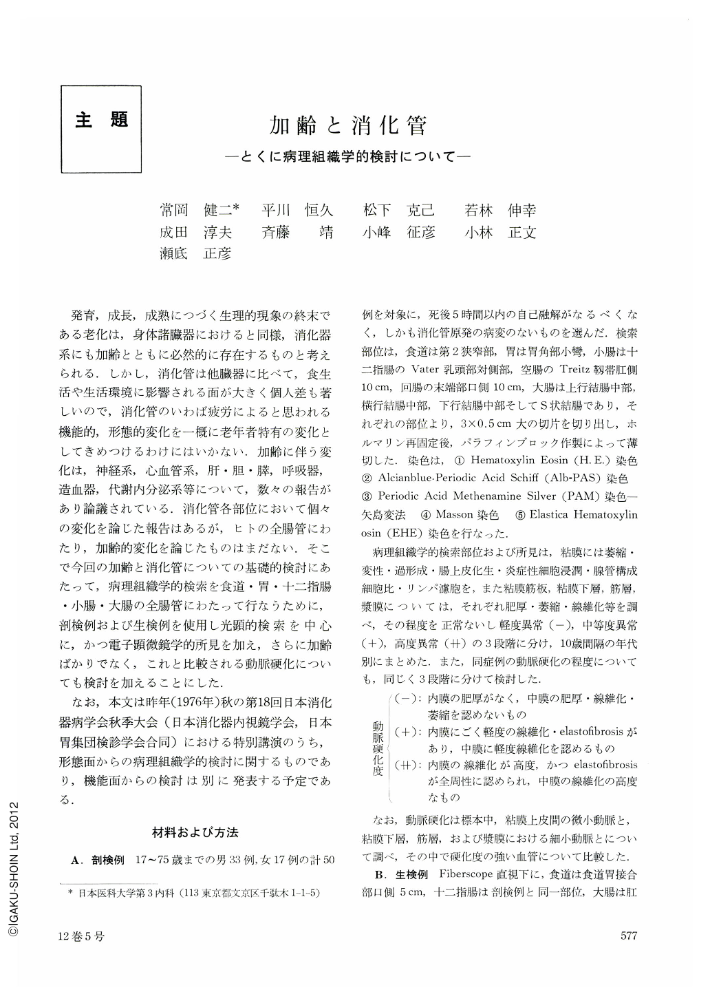
発育,成長,成熟につづく生理的現象の終末である老化は,身体諸臓器におけると同様,消化器系にも加齢とともに必然的に存在するものと考えられる.しかし,消化管は他臓器に比べて,食生活や生活環境に影響される面が大きく個人差も著しいので,消化管のいわば疲労によると思われる機能的,形態的変化を一概に老年者特有の変化としてきめつけるわけにはいかない.加齢に伴う変化は,神経系,心血管系,肝・胆・膵,呼吸器,造血器,代謝内分泌系等について,数々の報告があり論議されている.消化管各部位において個々の変化を論じた報告はあるが,ヒトの全腸管にわたり,加齢的変化を論じたものはまだない.そこで今回の加齢と消化管についての基礎的検討にあたって,病理組織学的検索を食道・胃・十二指腸・小腸・大腸の全腸管にわたって行なうために,剖検例および生検例を使用し光顕的検索を中心に,かつ電子顕微鏡学的所見を加え,さらに加齢ばかりでなく,これと比較される動脈硬化についても検討を加えることにした.
なお,本文は昨年(1976年)秋の第18回日本消化器病学会秋季大会(目本消化器内視鏡学会,日本胃集団検診学会合同)における特別講演のうち,形態面からの病理組織学的検討に関するものであり,機能面からの検討は別に発表する予定である.
In order to clarify the histopathological changes of the digestive tract in aging, we selected 50 autopsy cases soon after death without a primary pathology of the digestive tract and made light microscopic observations in one section each from the esophagus, stomach, duodenum, jejunum, ileum and four sections from the colon. Biopsy materials of the esophagus, duodenum and colon in cases without a digestive tract disease were also taken from approximately corresponding sites under fiberscopic observation and histologically examined. Futhermore, observations of biopsy materials by transmission as well as scanning electron microscopes and the semiquantitative analysis of DNA in the nuclei were carried out. And comparative studies were made in different age groups and arteriosclerotic groups.
The following results were obtained.
1) Hyperplasia of the esophageal mucosa in patients in their forties and fifties and slight atrophy of it in older age groups were observed. Atrophy of the muscular coat was also seen with aging. These findings were essentially similar in autopsy as well as in biops ycases. In scanning electron microscopic observations, linear surface structures became thick, coarse and irregular in cases of mucosal hyperplasia.
2) Intestinal metaplasia of the stomach became more extensive with aging and the goblet cells increased in number. There was also a tendency to increase in infiltration of inflammatory cells with aging. However, no definitive correlation between these findings were demonstrated in different arteriosclerotic groups.
3) The duodenum showed only a slight tendency to increase in the atrophy of the mucosal epithelium and also in the infiltration of inflammatory cells with aging. Wrinkling patterns of the duodenal villi running longitudinally as well as crosswise on scanning electron microscope seemed rather smooth in cases showing mucosal atrophy.
4) Atrophy became prominent in the mucosa and the muscular coat of the jejunum and the ileum with aging. Arteriosclerotic changes and also fibrosis gradually increased parallel to aging.
5) In the colon, atrophy of the mucosal epithelium as well as muscular coat increased with aging and was associated with the increase in number of the granular cells in the bottom of the crypts. On scanning electron microscopic findings, the colonic crypts in cases showing a mucosal atrophy became shallow and the goblet cells seemed to increase.
6) Arteriosclerotic changes were seen in persons over the age of 30 with some exceptions. They were more common and more severe with aging. The small arteries adjacent to the basement membrane of the epithelial cells showed essentially the same changes in different sites, which were also similar light-and electronmicroscopically.
7) The basement membrane of the mucosal epithelial cells was rather well-preserved on light-and electron microscopes and showed no difference in different age groups in spite of the presence of more or less atrophy of the mucosal epithelium with aging.
8) Several noticeable findings on scanning electron microscope to confirm light microscopic findings were obtained.
9) In the semiquantitative analysis of DNA in biopsy materials of the colonic mucosa, DNA decreased in volume with aging.

Copyright © 1977, Igaku-Shoin Ltd. All rights reserved.


