Japanese
English
- 有料閲覧
- Abstract 文献概要
- 1ページ目 Look Inside
十二指腸において,Vater乳頭部は,十二指腸球部についで病変の多い部位であり,特に膵胆道系との関連において重要である.内視鏡的にVater乳頭を観察した記録は,1966年Watoson1)らによるものが当初である.本邦においても,十二指腸鏡開発当初より,乳頭観察は大きな目標の一つであった.そのため,乳頭の観察には意欲的な努力が払われて来た.そして今日,ごく特殊なものを除けば,乳頭観察は全例に可能となった.また,観察のみではなく,生検,逆行性膵胆道造影が可能となり,乳頭部領域,膵胆道疾患の診断に極めて重要な情報を提供してくれる検査法として,臨床的にも次第に評価され,その位置が確立されつつある.
名称
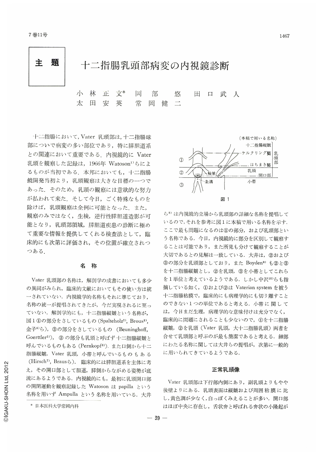
Vater乳頭部の名称は,解剖学の成書においても多少の異同がみられ,臨床的文献においてもその使い方は統一されていない.内視鏡学的名称もそれに準じており,名称の統一が提唱されてきたが,今だ実現されるに至っていない.解剖学的にも,十二指腸縦皺という名称が,図1①の部分をさしているもの(Spalteholz2),Brous3),金子4)ら),③の部分をさしているもの(Beuninghoff,Goerttler5)),②の部分も乳頭と呼ばず十二指腸縦皺と呼んでいるものもある(Pernkopf6)),また口側から十二指腸縦皺,Vater乳頭,小帯と呼んでいるものもある(Hirsch7),Brausら).臨床的には膵胆道系を主体に考え,その開口部として胆道,膵側からながめる姿勢が底流にあるようである.内視鏡的にも,最初に乳頭開口部の開閉運動を観察記録したWatosonはpapillaという名称を用いずAmpullaという名称を用いている.大井ら8)は内視鏡的立場から乳頭部の詳細な名称を提唱しているので,それを参考に図1に本稿で用いる名称を示す.ここで最も問題になるのは①の部分,および乳頭部という名称である.今日,内視鏡的に部分を区別して観察することは可能であり,また所見も分けて観察することが大切であるとの見解は一致している.大井は,②および③の部分を乳頭部としており,またBoyden9)も②と③を十二指腸縦皺とし,②を乳頭,③を小帯としてこれらを1単位と考えているようである.しかし中沢10)らも指摘している如く,①および②はVaterian systemを被う十二指腸粘膜で,臨床的にも病理学的にも切り離すことのできない1つの単位であると考える.小帯に関しては,今日まだ生理,病理学的な意味付けは充分でなく,臨床的に問題にされることも少ないので,①を十二指腸縦皺,②を乳頭(Vater乳頭,大十二指腸乳頭)両者を合せて乳頭部と呼ぶのが最も簡潔であると考える.細部にわたる名称に関しては大井らの提唱が,次第に一般的に用いられてきているようである.
Progress in duodenofiberscopic examination has not only made it easy to observe the region of the papilla of Vater but also made it possible to arrive at comprehensive diagnosis including biopsy and retrograde visualization of the pancreatic and biliary ducts. At present there still remains some confusion regarding the appropriate terms of this region.
Anatomy has not too standardized name for it, and clinically a common expression is employed. Endoscopic examination, depending solely on observation with the naked eye, needs a standardized, clear-cut nomenclature for it, as it is necessary to accurately describe findings observed. In this paper are used such names as the longitudinal fold, papilla and frenulum, and the longitudinal fold and papilla are collectively called as the region of the papilla of Vater.
Cancer in the papillary region is the most important lesion in this area. If radical operation be carried out at a proper time, prognosis would be much improved. As it is a lesion that permits easy access to endoscopy, correct diagnosis of cancer in this region is all the more important. Endoscopic findings of it are expressed more or less in such terms as swelling of the longitudinal fold, edematous protrusion of the papilla, its unevess, erosion, bleeding or tendency to it. Co-employement of biopsy and pancreatocholedochography is essential.
Papillitis is known as a benign stricture at the distal end of the common bile duct, but the site of the lesion belongs to the Vaterian system, and it is seen as change in the papillary region. Since it is a submucosal change, however, diagnosis is not always easy. Differention from cancer calls for more detailed investigation.
Choledocholithiasis is often accompanied with icterus, and it becomes an objective of duodenal examination, retrograde choledochography being the most effective method of diagnosis for it. Changes in the papilla are often slight, while protrusion of the longitudinal fold is mostly manifest.
Endoscopically papillary diskinesia is twofold: either the orifice remains open or its site becomes obscure owing to decreased function of the sphincter.
In parapapillary diverticulum we were unable to find out any change of the papillary region effected by it.
Together with endoscopy of the papillary region, pancreatocholedochography is most effective in the diagnosis of lesions in this area and in biliary and pancreatic ducts. Several cases are illustrated in this paper.
Copyright © 1972, Igaku-Shoin Ltd. All rights reserved.