Japanese
English
- 有料閲覧
- Abstract 文献概要
- 1ページ目 Look Inside
- サイト内被引用 Cited by
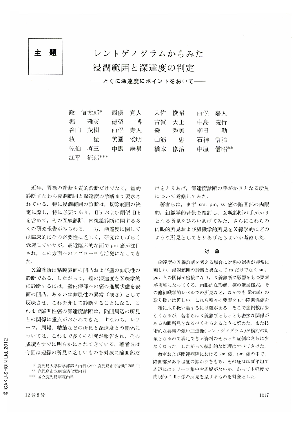
近年,胃癌の診断も質的診断だけでなく,量的診断すなわち浸潤範囲と深達度の診断まで要求されている.特に浸潤範囲の診断は,切除範囲の決定に際し,特に必要であり,Ⅱbおよび類似Ⅱbを含めて,そのX線診断,内視鏡診断に関する多くの研究報告がみられる.一方,深達度に関しては臨床的にその必要性に乏しく,研究はしばらく低迷していたが,最近臨床的な面でpm癌が注目され,この方面へのアプローチも活発になってきた.
X線診断は粘膜表面の凹凸および壁の伸展性の診断である.したがって,癌の深達度をX線学的に診断するには,壁内深部への癌の進展状態を表面の凹凸,あるいは伸展性の異常(硬さ)として反映させ,これを介して診断することになる.これまで陥凹性癌の深達度診断は,陥凹周辺の所見との関係に重点がおかれてきた.すなわち,レリーフ,周堤,結節などの所見と深達度との関係にっいては,これまで多くの研究が報告され,その成績もすでに明らかにされてきている.著者らは今回は辺縁の所見に乏しいものを対象に陥凹部だけをとりあげ,深達度診断の手がかりとなる所見について考察してみた.
There have been many reports about the findings around depressed areas of the stomach such as folds patterns and protrusions to estimate the invasion depth of depressed carcinoma. In this paper, based on detailed observations of the depressed area of a Ⅱc-lesion and two Ⅱc-like advanced cancers, a clue for the estimation of invasion depth in this area is discussed.
In addition to the thickened submucosa, the macroscopic and histological characteristics of sm-, pm-, and ss-carcinoma were deeper erosion in the mucosa, mucosal defect above the submucosa, its outcrop and so forth. Roentgenologically, these findings were recognizable in the resected specimens as follows: (1) The thickening of the submucosa was recognizable as a filling defect on compression pictures.
(2) Deeper erosion in the mucosa, mucosal defect above the thickened submucosa, and its outcrop were recognizable on double contrast radiographs as indistinct or disappeared mucosal pattern, and various mucosal pictures different from the pattern proper to each specimen.
Further study is necessary to make it clear how to recognize these findings in preoperative X-ray examination.

Copyright © 1977, Igaku-Shoin Ltd. All rights reserved.


