Japanese
English
- 有料閲覧
- Abstract 文献概要
- 1ページ目 Look Inside
症 例
患 者:瀬○ロ 71歳 ♂
特記すべき主訴はなく,ドックのために入院した.
家族歴:特記すべき事項なし.
既往歴:5年前下血があり,胃潰瘍として治療を行っている.
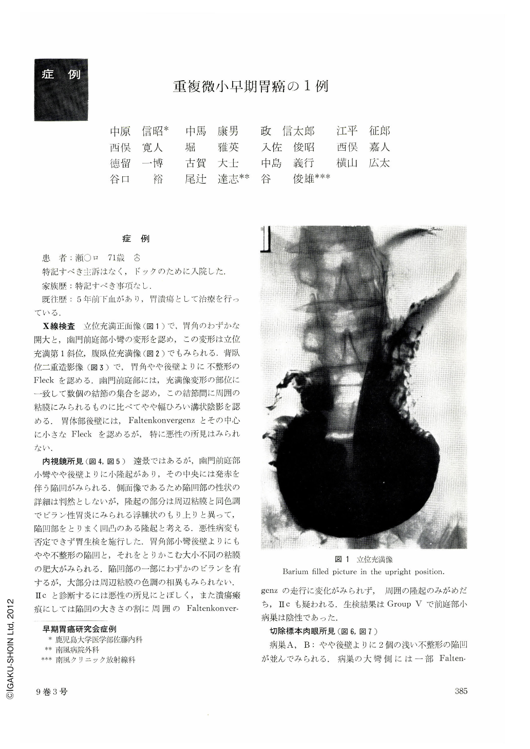
A 71 years old male visited our clinic for the purpose of a through physical checkup and, as a result, deformation was found by roentgenographic examination in the regions of angulus ventriculi and pyloric antrum. Double contrast examination revealed irregular “Fleck” in the posterior wall of the angulus ventriculi and a sulciform shadow within a granular shadow in the pyloric antrum region. Macroscopic observation showed similar finding, as shown by roentgenographic examination, of a shallow, irregular excavated lesion located in some measure to the posterior wall and of a sulciform ulcerative lesion with its mucosa being elevated located in a measure to the posterior wall of pyloric antrum. Both lesions were histologically adenocarcinoma tubulare.
The minute gastric cancer found in the pyloric antrum was 2×7 mm in size and infiltrated along macroscopically excavated lesion, but did not whole sulciform lesion area. In other words, though the minute gastric cancer was detected by x-ray examination in the form of a sulciform shadow, its sphere did not coincide with the sulciform shadow. The sulciform shadow was wider and denser and its margin more sharp when compared with “Fleck” in the non-infiltrated lesion.
Up to this time, in the field of roentgenographic examination much attention has been paid to reveal the resected specimen exactly. Except this, however, x-ray examination has another advantage of showing indentation of area, which may contribute to the diagnosis of the minute gastric cancer.

Copyright © 1974, Igaku-Shoin Ltd. All rights reserved.


