Japanese
English
- 有料閲覧
- Abstract 文献概要
- 1ページ目 Look Inside
- サイト内被引用 Cited by
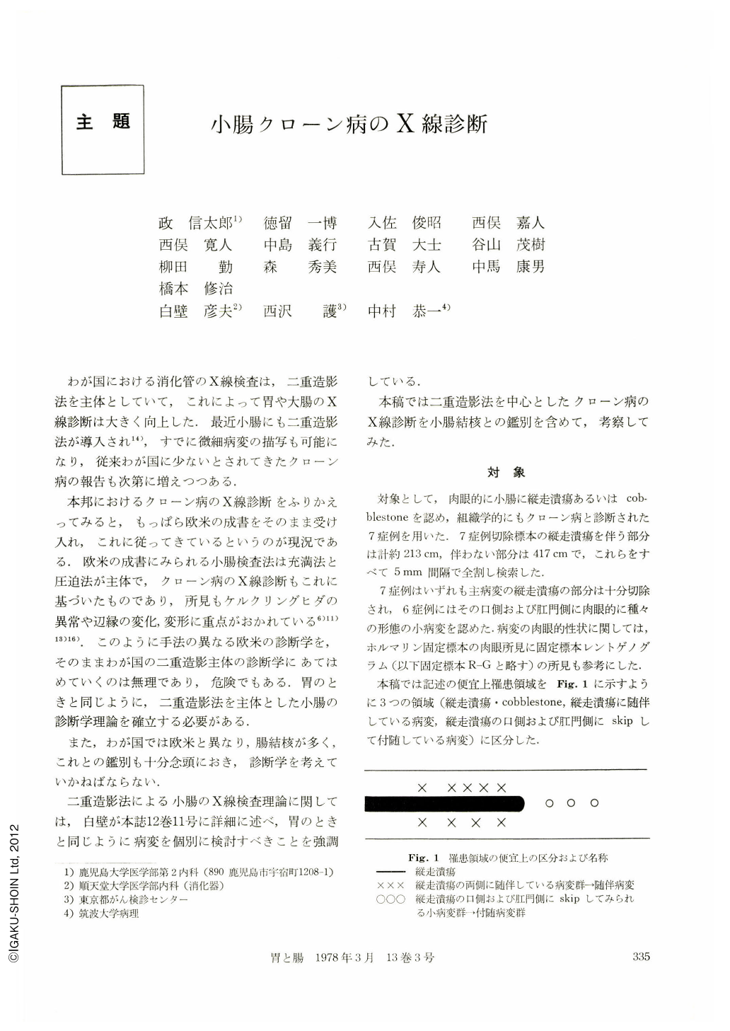
わが国における消化管のX線検査は,二重造影法を主体としていて,これによって胃や大腸のX線診断は大きく向上した.最近小腸にも二重造影法が導入され14),すでに微細病変の描写も可能になり,従来わが国に少ないとされてきたクローン病の報告も次第に増えつつある.
本邦におけるクローン病のX線診断をふりかえってみると,もっぱら欧米の成書をそのまま受け入れ,これに従ってきているというのが現況である.欧米の成書にみられる小腸検査法は充満法と圧迫法が主体で,クローン病のX線診断もこれに基づいたものであり,所見もケルクリングヒダの異常や辺縁の変化,変形に重点がおかれている6)11)13)16).このように手法の異なる欧米の診断学を,そのままわが国の二重造影主体の診断学にあてはめていくのは無理であり,危険でもある.胃のときと同じように,二重造影法を主体とした小腸の診断学理論を確立する必要がある.
Crohn's disease of the small intestine in 7 cases was studied roentgenographically, mainly by means of double contrast method. All of those cases had longitudinal ulcers and/or cobblestone appearance and were pathologically diagnosed as Crohn's disease. For descriptive purpose, the lesions seen in Crohn's disease of the small intestine were divided into macroscopically three resions:
1) Area of longitudinal ulcer and cobblestone appearance.
2) Area associated with the longitudinal ulcers.
3) Skip lesions on the oral side of the longitudinal ulcers.
On double contrast examination lesions on the mesenteric borders of the intestine were usually demonstrated as en-face views in consequence of rotation or distortion of the small intestine, which is favor of making diagnosis of Crohn's disease.
It is to be mentioned that not only longitudinal ulcers and ‘cobblestone’ appearance but other small lesions were easily demonstrated as direct sign on double contrast radiograph. However, lesions associated with severe deformity, especially those in the stenosed parts of the intestine, were not demonstrated clearly. Therefore, diagnosis of the lesion associated with severe stenosis should be made according to type and severity of deformity of the affected parts.
Accurate diagnosis of the features of each lesion is necessary to differentiate it from intestinal tuberculosis which is often seen in Japan.
Relation between macroscopic features of lesion and type and severity of deformity was studied and following results were obtained.
1) Severe stenosis of the affected part was closely associated with transverse lesion in most cases.
2) Concentric stenosis of the intestine seen in Crohn's disease was difficult to be differentiated from ‘annular’ stenosis seen on X-ray findings of girdle ulcer of intestinal tuberculosis. Analysis of X-ray findings of the stenotic segment of the intestine, however, enabled us to differentiate them in most cases.
3) The process toward advanced stage of Crohn's disease will be made clear by detecting small changes of lesions in the small intestine in vivo.
Macroscopically, the limits of lesion is well-defined in Crohn's disease. A normal-looking mucosa (Kerkring's folds, tips of converging folds) seems to be adjacent to the margin of the lesion.
Therefore, it is important to differentiate from the girdle ulcer of small intestinal tuberculosis.

Copyright © 1978, Igaku-Shoin Ltd. All rights reserved.


