Japanese
English
- 有料閲覧
- Abstract 文献概要
- 1ページ目 Look Inside
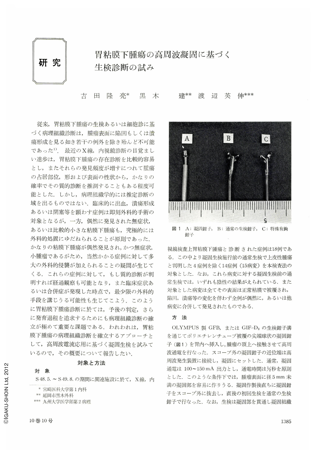
従来,胃粘膜下腫瘍の生検あるいは細胞診に基づく病理組織診断は,腫瘤表面に陥凹もしくは潰瘍形成を見る如き若干の例外を除き殆んど不可能であった1).最近のX線,内視鏡診断の目覚ましい進歩は,胃粘膜下腫瘍の存在診断を比較的容易とし,またそれらの発見頻度が増すにつれて腫瘍の占居部位,形および表面の性状から,かなりの確率でその質的診断を推測することもある程度可能とした.しかし,病理組織学的には推定診断の域を出るものではない.臨床的に出血,潰瘍形成あるいは閉塞等を顕わす症例は即刻外科的手術の対象となるが,一方,偶然に発見された無症状,あるいは比較的小さな粘膜下腫瘍も,究極的には外科的処置にゆだねられることが原則であった.かなりの粘膜下腫瘍が偶然発見され,かつ無症状,小腫瘤であるがため,当然かかる症例に対して多大の外科的侵襲が加えられることの疑問が生じてくる.これらの症例に対して,もし質的診断が判明すれば経過観察も可能となり,また臨床症状あるいは合併症が発現した時点で,最少限の外科的手段を講じうる可能性も生じてこよう.このように胃粘膜下腫瘍診断に於ては,予後の判定,さらに発育過程を追求するためにも病理組織診断の確立が極めて重要な課題である.われわれは,胃粘膜下腫瘍の病理組織診断を確立するアプローチとして,高周波電流応用に基づく凝固生検を試みているので,その概要について報告したい.
We introduced electrocoagulation biopsy as a new method for making histopathological diagnosis of submucosal tumor of the stomach which has no ulceration over it, since the diagnosis of histologic nature of submucosal tumor has been extremely difficult by routine biopsy technic regardless of size. Electrocoagulation biopsy was applied to 14 cases (15 tumors) which were diagnosed to be submucosal tumor of the stomach by upper gastrointestinal series, gastroscopy and conventional biopsy. Histopathological diagnosis was obtained by this procedure in 7 cases; 2 leiomyomas, 2 lipomas, 1 ieiomyoblastoma, 1 aberrant pancreas and 1 schistosomiasis. In addition, 2 cases were also strongly suspected to be leiomyoma in histology. Positive electrocoagulation biopsy was, therefore, obtained in 9 out of 14 cases. In terms of the size of the tumor, histologic nature was documented in all 8 tumors but one more than 2 cm in greatest diameter. On the contrary, there was no positive result obtained in the tumors less than 1.0 cm in size except for one case. The first biopsy immediately after electrocoagulation failed to demonstrate tumor tissue in all 9 case but one. However, subsequent biopsies 2 to 3 days after electrocoagulation have verified histologic nature in all 9 cases. Necrotic changes and regenerative granulation tissues due to electrocoagulation have been the factors which made histological interpretation of biopsy specimen difficult, especially in cases of negative result. Difference in consistency of the tumor has also been an embarrassing factor in making histological diagnosis due to inability of obtaining tumor tissue, i.e. in leiomyoma. Introduction of a new special biopsy forceps will be strongly expected in order to overcome negative biopsy resulting from tissue reaction due to electrocoagulation and difference in consistency of the tumor.
Our materials consisting of 14 cases of submucosal tumor of the stomach were all found incidentally by conventional examinations, and except for an operated case of leiomyoblastoma, they are now being under strict observation because they remain asymptomatic and the tumors are small in size.

Copyright © 1975, Igaku-Shoin Ltd. All rights reserved.


