Japanese
English
- 有料閲覧
- Abstract 文献概要
- 1ページ目 Look Inside
早期胃癌の診断に関しては,その周辺粘膜の微細構造についてのX線及び内視鏡像の検討から,1cm以下の病変を発見することも可能となったが,一方では胃の大きな病変が,その大きさゆえに癌と誤診されるという症例も散見されるようである.われわれは,術前胃幽門前庭部の進行胃癌と診断し,切除胃標本の組織学的検索から,胃幽門部粘膜の広範な幽門腺肥厚を示したMénétrier1)病と考えられる例で,悪性所見の認められなかった症例を経験したので,文献的考察を加え報告する.
症例
患 者:63歳,男子,会社役員.
主 訴:上腹部鈍痛.
家族歴,生活歴,既往歴に特記すべきことはない.
現病歴:入院2週間前より鈍い上腹部痛を感じるようになった.悪心,嘔吐,胸やけなどはなく,食欲も正常であった.全身倦怠感がつよく,疲れやすいので某医を受診し,胃X線検査を受け,胃に病変を指摘され手術をすすめられたので当科を訪れた.現在上腹部の鈍痛があるのみである.
The patient : a man 63 years of age. Chief complaint : dull pain in the upper part of the abdomen. Since two weeks before admission he had begun to complain of dull pain in the upper abdomen in addition to general lassitude. When he had been examined elsewhere with x-ray a lesion was found in the stomach. He was then advised to undergo surgical intervention in our hospital.
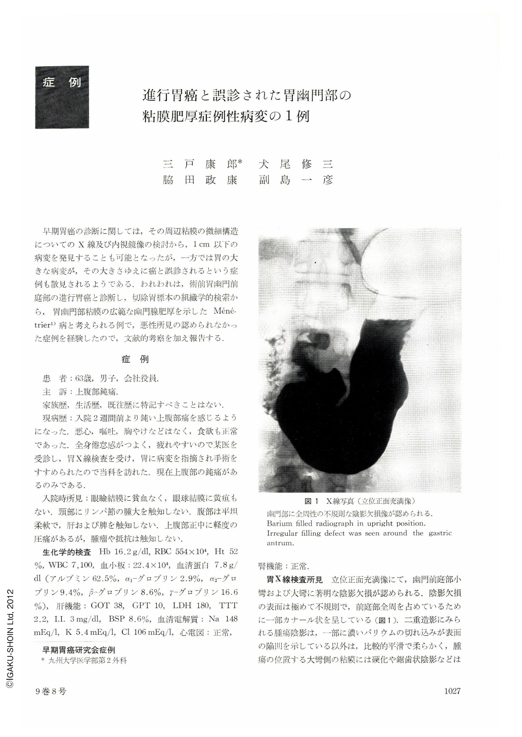
While upright barium-filled picture revealed a shadow defect of irregular shape in the pyloric antrum extending to the whole circumference of the wall, the shadow of a tumor in double contrast view was seemingly of softer outline with smooth surface. The surrounding mucosa was neither rigid nor irregular in pattern. The whole picture was a well depicted protrusion, smooth-surfaced and without ulceration. Nonetheless, the mucosa directly around the tumor looked slightly rigid and there were some changes as well suggesting malignant infiltration through the pyloric ring into the duodenal cap. These findings were suggestive of advanced carcinoma of a protruding variety.
Endoscopy also revealed a mucosal protrusion with surface erosions that looked soft and extended all around the pyloric antrum. But seen tangentically from the oral side, the entire aspect of the tumor was hidden from view and the presence or absence of ulceration could not be determined. We then made a tentative diagnosis of either cancer or malignant submucosal tumor of the stomach, with only its oral aspect exposed to the view.
Resected stomach showed a wide, plateau-like protrusion with relatively localized margins on the oral side, measuring 6 . 5 X 9. 0 X 1. 5 cm, extending over the whole circumference of the pyloric antrum. There were groove-like depressions on the surface running in an irregular fashion, suggesting shallow interrugal sulci, some of them looking at first sight like cerebral convolutions. Although erosive spots were seen scattered about, no ulceration was in evidence.
Histolgoically, the mucosa over the protrusion was thickened on account of striking hyperplasia of the pyloric glands. Muscle fibers in the muscularis mucosae increased in number, rising upward among the hyperplastic glands. Foveolar hyperplasia or cystic formation of the glands was hardly recognized. Not a malignant finding was thus in sight. These findings were believed to belong to the category of “ Polyadenomes en nappe” according to the original report of P. Menetrier. Usually the so-called Ménétrier's disease arises on the side of the greater curvature. " Polyadenomes en nappe " localized within the pyloric region have been reported so far in 3 cases only, including the reports by Nakamura and Martini.
Copyright © 1974, Igaku-Shoin Ltd. All rights reserved.