Japanese
English
- 有料閲覧
- Abstract 文献概要
- 1ページ目 Look Inside
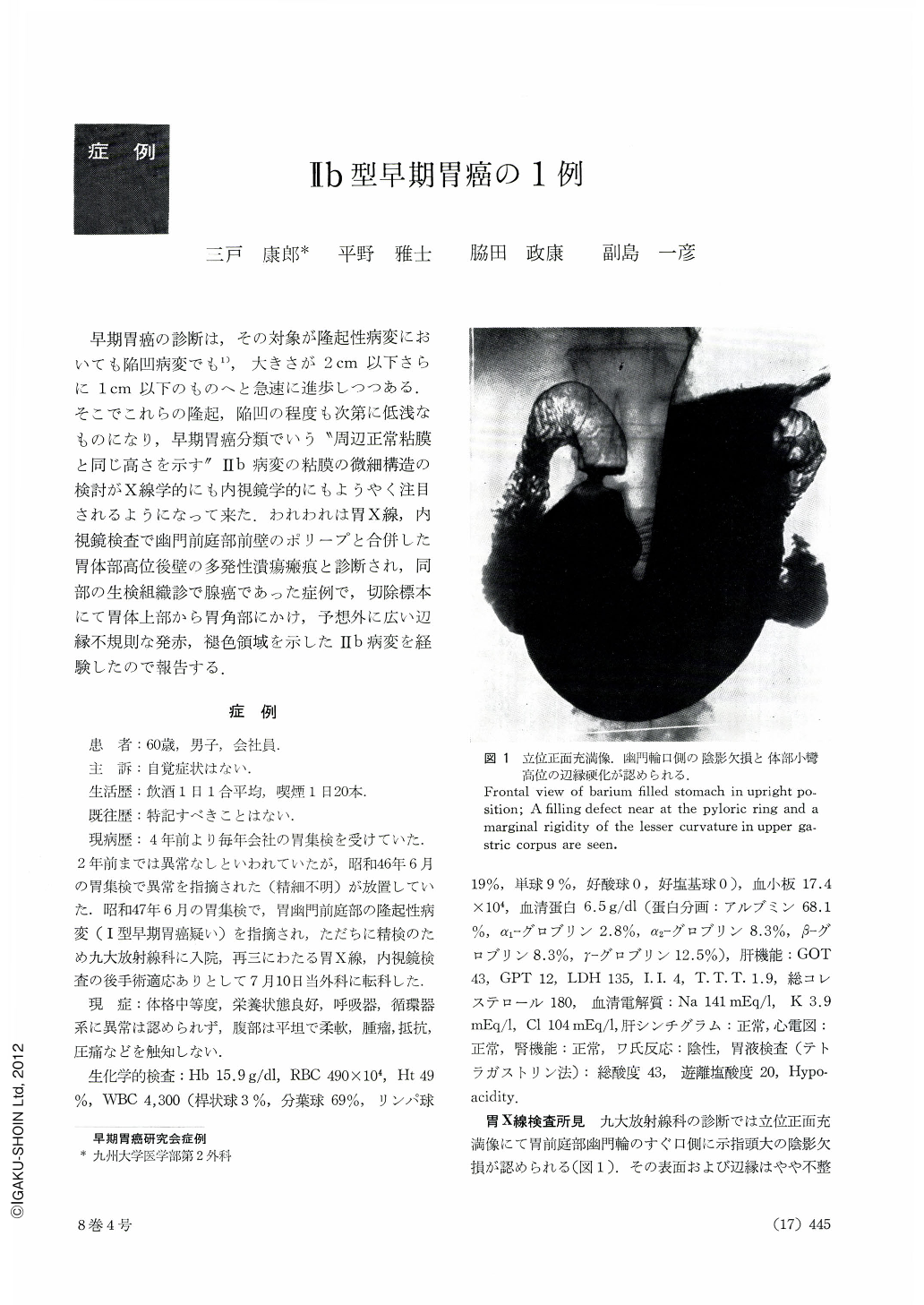
早期胃癌の診断は,その対象が隆起性病変においても陥凹病変でも1),大きさが2cm以下さらに1cm以下のものへと急速に進歩しつつある.そこでこれらの隆起,陥凹の程度も次第に低浅なものになり,早期胃癌分類でいう“周辺正常粘膜と同じ高さを示す”Ⅱb病変の粘膜の微細構造の検討がX線学的にも内視鏡学的にもようやく注目されるようになって来た.われわれは胃X線,内視鏡検査で幽門前庭部前壁のポリープと合併した胃体部高位後壁の多発性潰瘍瘢痕と診断され,同部の生検組織診で腺癌であった症例で,切除標本にて胃体上部から胃角部にかけ,予想外に広い辺縁不規則な発赤,褪色領域を示したHb病変を経験したので報告する.
A man aged 60 had undergone annual mass screening of the stomach at his place of work since four years before. Two years later an abnormality was detected in his stomach, but he neglected to seek medical help. At a mass survey in June 1971 a protruding lesion (suspicion of type Ⅰ early cancer) was found in the stomach, and this time he was hospitalized for thorough check-up in the Department of Radiology, College of Medicine, Kyushu University. After examined with x-ray and endoscopy, he was referred to our Department of Surgery with an indication for surgical intervention. Preoperative x-ray study revealed, besides a broad-based protruding lesion of mushroom shape in the greater curvature side of the antrum, an abnormal area on the posterior wall near the angulus. Thin barium flecks adheared to it and areae gastricae were obliterated. This area was surrounded by a strip of areae gastricae, irregular both in shape and size. Towards the obliterated areae gastricae was seen irregular rugal convergency, and the contour of the lesser curvature was rigid. Granular elevation was also seen on the oral side. It was head to distinguish from a Ⅱc lesion, but tentatively a diagnosis of mutiple ulcer scars was made Endoscopy also revealed there spots of white coat and erosion scattered about, and the diagnosis was multiple ulcers. However, biopsy disclosed cells of well differentiated adenocarcinoma.
Besides the mushroom-shaped polyp already mentioned on the greater curvature of the antrum, resected stomach showed flat area, 9.5 cm in the greatest diameter, extending from the upper part of the body right down to the level of the angle, with the lesser curvature in the center. The areae gastricae were obliterated, dotted with irregular engorgement. Histologically, it was well differentiated adenocarcinoma with flat margins and limited within the mucosal layer. There was no lymph node involvement. What was most striking in this case was that it was a typical Ⅱb lesion, to be distinguished from the normal mucosa barely by gross observation of irregular reddening. In cut sections as well, it belonged to the flat B group (Nishizawa).

Copyright © 1973, Igaku-Shoin Ltd. All rights reserved.


