Japanese
English
- 有料閲覧
- Abstract 文献概要
- 1ページ目 Look Inside
胃において急性に発生する病変の中で,臨床的に最近注目されているものに,高木ら1)の報告した胃幽門前庭部の急性対称性潰瘍と,唐沢ら2)の検討した出血性びらんがある.高木はこの急性対称性潰瘍も出血性びらんも胃前庭部の平滑筋痙攣が急激に,かつ異常に強く発生して血行障害を起したために生じたもので,発症要因の程度により,時に潰瘍を作り,時にびらんを生じるものと推定している.これらの疾患は急激に発症するので,発症直後に診療する機会の多い実地医家にとり殊に重要なものである.昭和42年7月から昭和48年12月までに,私共は急性対称性潰瘍と出血性びらんとを合せて25例経験した.この経験を基に,両病変の臨床的事項,X線及び内視鏡所見についてのべ,両者の関係について検討を加えてみたい.
症例
〔症例1〕患者:17歳,男子高校生.
前庭部の急性対称性潰瘍.特別の既往なし.
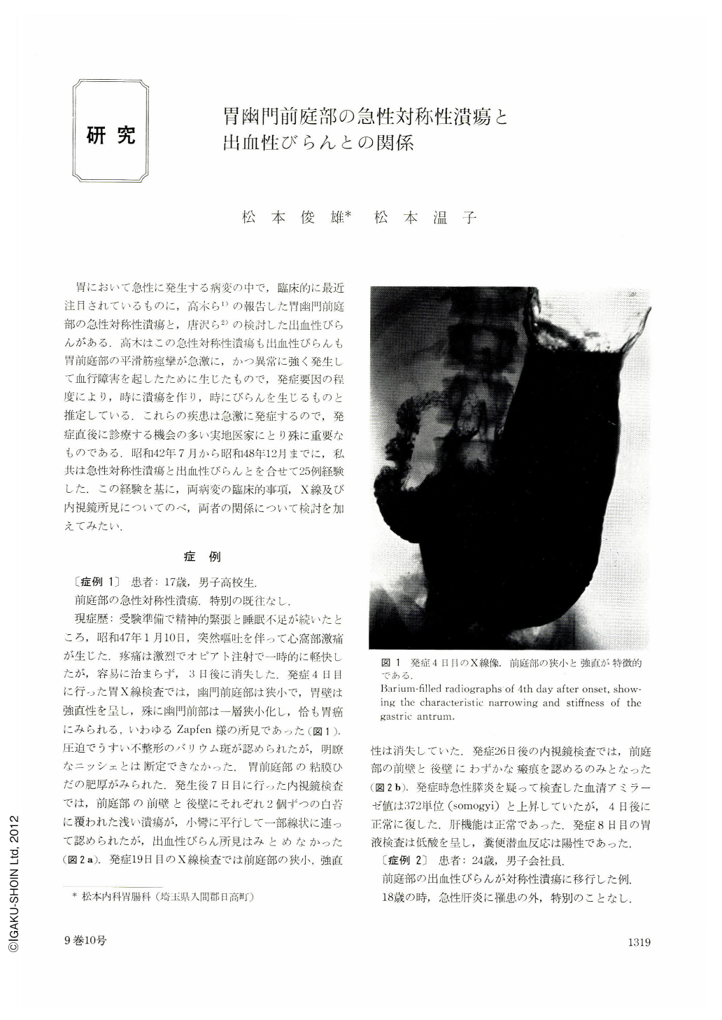
現症歴:受験準備で精神的緊張と睡眠不足が続いたところ,昭和47年1月10日,突然嘔吐を伴って心窩部激痛が生じた,とう痛は激烈でオピアト注射で一時的に軽快したが,容易に治まらず,3日後に消失した.発症4日目に行った胃X線検査では,幽門前庭部は狭小で,胃壁は強直性を呈し,殊に幽門前部は一層狭小化し,恰も胃癌にみられる,いわゆるZapfen様の所見であった(図1).圧迫でうすい不整形のバリウム斑が認められたが,明瞭なニッシェとは断定できなかった.胃前庭部の粘膜ひだの肥厚がみられた.発生後7日目に行った内視鏡検査では,前庭部の前壁と後壁にそれぞれ2個ずつの白苔に覆われた浅い潰瘍が,小彎に平行して一部線状に連って認められたが,出血性びらん所見はみとめなかった(図2a).発症19日目のX線検査では前庭部の狭小,強直性は消失していた.発症26日後の内視鏡検査では,前庭部の前壁と後壁にわずかな瘢痕を認めるのみとなった(図2b).発症時急性膵炎を疑って検査した血清アミラーゼ値は372単位(somogyi)と上昇していたが,4目後に正常に復した.肝機能は正常であった,発症8日目の胃液検査は低酸を呈し,糞便潜血反応は陽性であった.
There are some common features between acute symmetric ulceration and haemorrhagic erosion of the antral portion of the stomach in the clinical, roentogenological and endoscopic aspects.
They attack most frequently the young persons, even though all ages in both sexes are affected. Their onset begins suddenly with severe epigastric pain accompanied with nausea and vomiting in most cases. Such symptoms are apt to cease gradually after 2 to 3 days. The gastric acid tends to show hypoacidity. The occult blood test in the stool is always positive, but no melena has been recognised.
The x-ray film pictures taken as early as possible after the onset always show the characteristic narrowing and the increased regidity with a slight degree of irregularity of the gastric wall of the antrum, which remined us of the changes due to gastric cancer, especially of scirrhous type, but these findings usually disappear after short duration of time. It is rather difficult to know correctly the presence of ulcer niches or their natures from the x-ray films of this stage.
The initial findings of endoscopy carried out at the earlier stage reveal either haemorrhagic erosions only or symmetric ulcerations mixed with sporadically scattered erosions around them, all of which are covered with black or reddish coating. The small-sized erosion has a tendency to disappear in a few days, but the greater-sized erosion and ulceration change at all times into acute symmetric ulceration with a white coating. Most of acute symmetric ulceration are said to be Ul-Ⅱ in depth histologically. Then, we regard it as appropriate to think that they are essentially the same kind of ulcerative disorder, though there is slight difference in the grade of severity of symptoms and clinical findings between them.
Moreover, we have experienced three special cases the first case had haemorrhagic erosions at the second portion of duodenum, the second case was transiently accompanied with an elevated serum amylase level, and the final case was concomitant with transiently increased levels of serum transaminase, alkaliphosphatase and amylase without showing any organic disturbances of the bile duct system and pancreas. If these clinical and laboratory findings are not accidentaly combined, there may be a possibility of the involvement of the causing factor of this disease not only to the stomach but also to the duodenum. Judging from the above-mentioned facts, this acute lesion seems to be different from the so-called gastric peptic ulcer in the etiologic mechanism. Though this disease is easily cured in a relatively short time, it is important in the practice of medicine, because it needs often be differentiated, firstly, from acute abdomen such as acute pancreatitis and gallstone attack in its initial symptoms; and secondly from gastric cancer in its roentgenologic and endoscopic pictures taken at the beginning stage; and finally at times from Ⅱc or Ⅱa early cancer when this disease is in the stage of acute symmetric ulcer or its scar.

Copyright © 1974, Igaku-Shoin Ltd. All rights reserved.


