Japanese
English
- 有料閲覧
- Abstract 文献概要
- 1ページ目 Look Inside
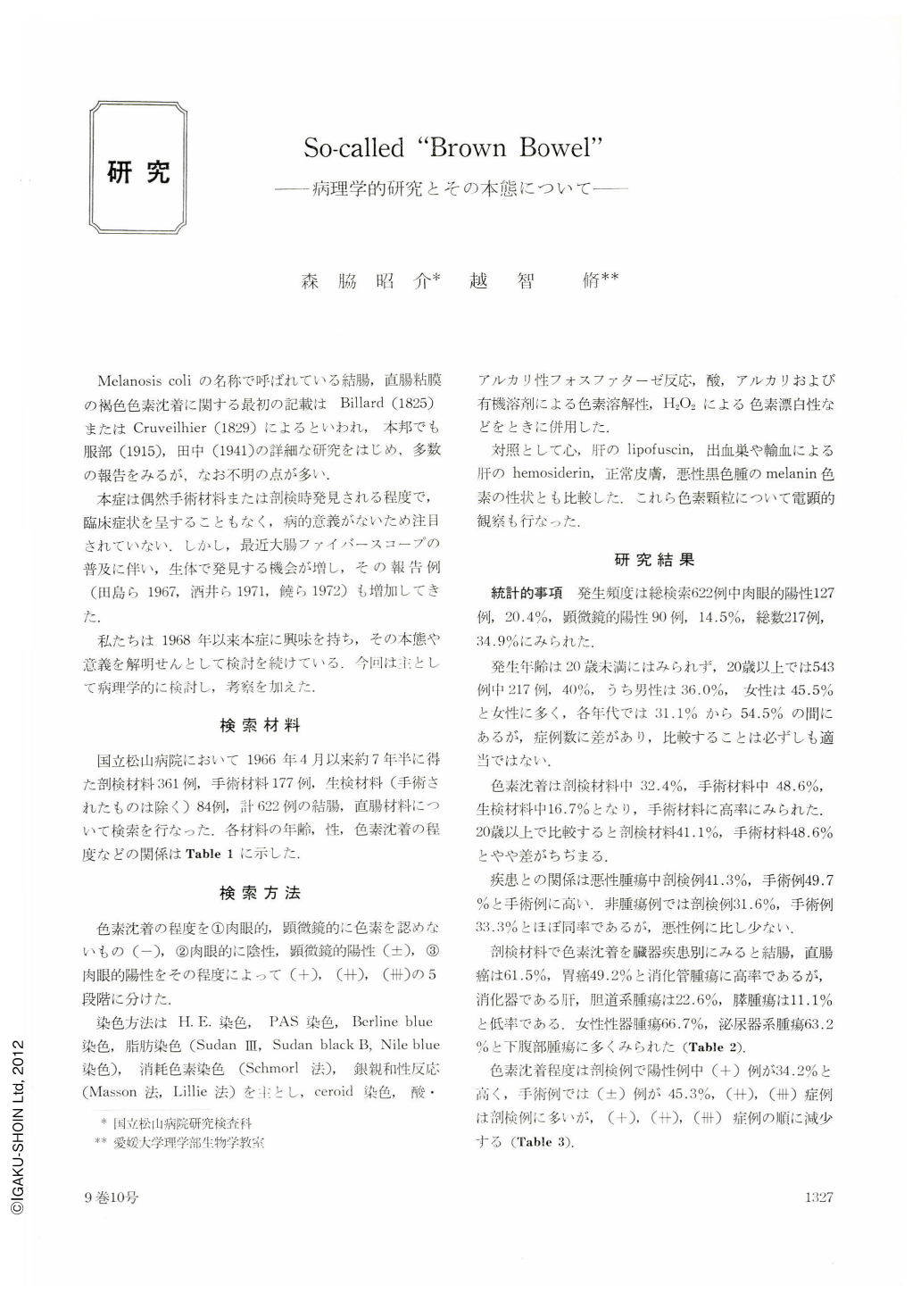
Melanosis coliの名称で呼ばれている結腸,直腸粘膜の褐色色素沈着に関する最初の記載はBillard(1825)またはCruveilhier(1829)によるといわれ,本邦でも服部(1915),田中(1941)の詳細な研究をはじめ,多数の報告をみるが,なお不明の点が多い.
本症は偶然手術材料または剖検時発見される程度で,臨床症状を呈することもなく,病的意義がないため注目されていない.しかし,最近大腸ファイバースコープの普及に伴い,生体で発見する機会が増し,その報告例(田島ら1967,酒井ら1971,饒ら1972)も増加してき
た.
The brown pigmentation of the recto-colonic mucosa called melanosis coli is not a rare condition, and numerous clinical and pathological studies have been reported, but the nature and significance of the pigments remains to be elucidated.
We have studied the clinico-pathologic, especially histochemic and electron-microscopic observation of the large bowel during a period of seven years and a half in our hospital.
The incidence of deposition of brown pigments in the human large bowel were found in 217 cases (34.9%) of 622 materials, -117 in 361autopsies, 86 in 177 operations and 14 in 84 biopsies. The frequency is higher in lower abdominal tumors-female sex organs (66.7%), urinary organs (63.2%) and large bowel (61.5%).
On light microscopic observations, the pigments were revealed to be similar to the lipofuscin in various staining. Electron-microscopically, the pigments differed from lipofuscin, hemosiderin and melanotic pigments. The pigments-conteining cells-phagcytes -were found in the mucosa and submucosa of the large bowel. The pigments changed from autophagosomes to autolysosomes and finally to residual bodies in phagocytes. Regarding the pigmentation we consider that chronic static contents of digestive tract are absorbed from the surface of the superficial lining cells, and then they are transmitted to the phagocytes in the tunica mucosa as hetrophagosomes. However, our present observation could not reveal the transforming process of pre-pigmented materials in epithelial cells.
One of the most important causes of pigmentation is chronic stasis of ingested materials in the digestive tract in various conditions.
The processes of pigmentations are related with lysosomes and a condition of lysosomal storage lesions.

Copyright © 1974, Igaku-Shoin Ltd. All rights reserved.


