Japanese
English
- 有料閲覧
- Abstract 文献概要
- 1ページ目 Look Inside
- サイト内被引用 Cited by
はじめに
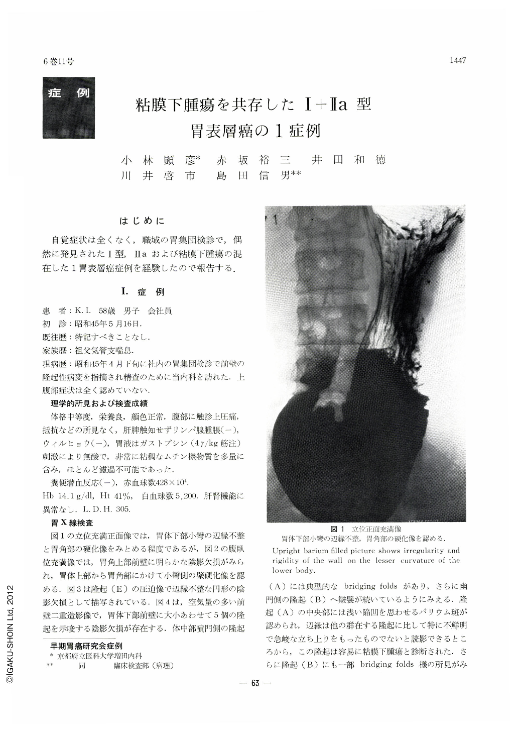
自覚症状は全くなく,職域の胃集団検診で,偶然に発見されたⅠ型,Ⅱaおよび粘膜下腫瘍の混在した1胃表層癌症例を経験したので報告する.
Superficial carcinoma of the stomach was diagnosed in a 58-year-old male, a company employe, on the basis of x-ray and endoscopy findings after he had been found to harbor an abnormality in his stomach at a gastric mass survey. He had no subjective symptoms to complain of at the time of thorough check-up. X-ray revealed a cluster of protruding lesions on the anterior wall of the lower body together with a submucosal tumor with typical bridging folds on the oral side. These protrusions partly looked like submucosal tumors but at the same time they might well have been suspected either as cancoma or sarcoma. By endoscopy they were observed as four protrusions with marked epithelial change belonging to type Ⅱ or Ⅲ as classified by Yamada. A submucosal tumor was also seen on their oral side. Histologically, these protruding lesions proved all to be adenocarcinoma muconodulare producing much mucus. Cancer infiltration had spread within the submucosal layer which contained large deposits of mucous substance within it, thereby forming an elevation above the mucosal surface. The irregular mucosal area surrounded by these protrusions was infiltrated with cells of adenocarcinoma tubulare, linking all of them together by cancer infiltration. The whole picture was superficial carcinoma sm degree of depth invasion. The reason why they looked partly like submucosal tumors was probably because cancer nests were not exposed around the bases of these protrusions but were only seen at their tips.
An inference on a development pattern of gastric cancer has also been attempted from the fact that atypical cells partly very close to cancer cells were found in the submucosal tumor in the oral side.

Copyright © 1971, Igaku-Shoin Ltd. All rights reserved.


