Japanese
English
- 有料閲覧
- Abstract 文献概要
- 1ページ目 Look Inside
はじめに
1888年Menetrierは胃の粘膜が胃腺の肥大によって限局性,もしくは瀰漫性に大脳回転を思わせる程に発達し,特異な巨大皺襞を呈する良性の疾患に病理組織学的な考察を加え,polyadenomesen nappeという名称を与えて報告した1).今日,Menetrier病といわれるのがこれである.しかし,本症には20に近い同義語2)~5)があり,臨床的概念および病理組織学的本態は未だ確立しているとはいい難い.
筆者らの症例は文献的考察から本症に該当するが,臨床診断学的ならびに病理組織学的に興味ある所見を呈したので,本症をめぐる問題点に若干の考察を加えて報告する.
A 51-year-old male, suffering from occasional epigastralgia for the past 15 years, visited the Author's clinic with an additional complaint of bouts of vomiting since two years before.
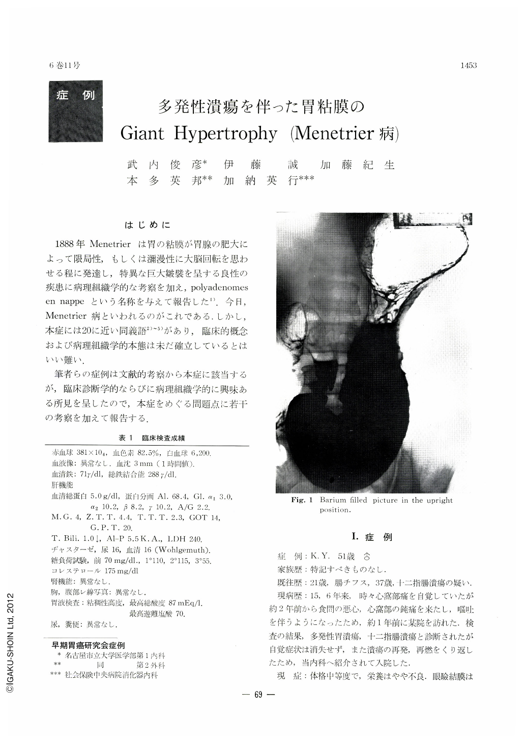
X-ray and endoscopy revealed the rugal pattern of the gastric mucosa except for the antrum replaced by hypertrophied folds, which were tortuous and perpentine, some lobulated or shortened, presenting as a whole a peculiar picture as if they were clusters of independent polypoid lesions. Multiple erosions and puckered ulcers were also seen between those broadened folds. However, distensibility of the gastric wall was well retained and the color tone of the mucosal surface, slightly more glossy without mucous substance, was almost normal. An ulcer was observed in the duodenal bulb as well.
Clinical examinations revealed hypoproteinemia, raised A/G ratio and decrease in serum iron. The gastric juice showed hyperacidity. Biopsy specimens also pointed to chronic gastritis and ulcers.
These findings led the author to the diagnosis of a peculiar type of gastritis associated with ulcers, ulcer scars and an ulcer in the duodenum.
Because the patient's complaints, persistent over 15 years, and his nutritional condition did not improve as was expected under medical treatment, with ulcers recurring or starting afresh, it was finally decided to perform total gastrectomy.
The resected stomach was of normal consistency. Hypertrophied folds in the opened stomach looked like a mass of worms. Multiple erosions and puckered ulcers were recognized in between.
Microscopic examination of the thickened gastric folds revealed mostly hyperplasia of the surface epithelium and glands, with normal proportion of chief, mucous and parietal cells retained. Inflammation of the interstitial tissue and the submucosa was slight, and the muscularis mucosae was smooth and of normal thickness. However, some broadened folds presented a picture of atrophic, hyperplastic gastritis. Between these folds were seen a number of erosions and ulcers with small converging folds. No malignant finding could be recognized in the entire extent of the gastric mucosa. The patient has been showing satisfactory progress toward recovery.
Although this case comes under Menetrier's disease, one presenting such peculiar features, clinically and pathologically, has not been reported hitherto in its literature.

Copyright © 1971, Igaku-Shoin Ltd. All rights reserved.


