Japanese
English
- 有料閲覧
- Abstract 文献概要
- 1ページ目 Look Inside
Ⅰ.はじめに
同一の基部より茎が樹枝状に分岐し,更に多数の腫瘤に分かれ,珊瑚状を呈した比較的まれな形状の胃ポリープと,その附近に平盤状のⅡa型早期胃癌が共存し,又門脈圧亢進による静脈瘤も合併し,複雑な所見を示した1症例を経験したので報告する.
This is a report of four protruding lesions co-existent in one stomach. The first lesion was a rare type of pedunclated polyp, having several stalks like those of a tree, which were further divided into smaller tumors, resembling a coral in its outward look. In its neighborhood was also found a small sessile polyp. The third lesion belonged to a Type Ⅱa early gastric cancer, the fourth being varices in the cardiac region due to portal hypertention.
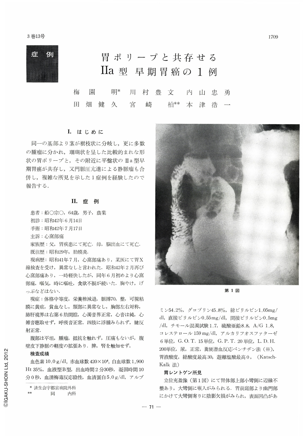
X-ray studies in barium filled stomach in upright position revealed a filling defect in the greater curvature side from the antrum down to the pyrolus and partly through the latter onto a part of the duodenal bulb. In the prone and supine double contrast studies, a lesion was visualized, extending from the antrum to the pyrolus, of irregular outline with marked surface unevenness, suggesting conglomeration of many small tumors there. Its location was not fixed but flexible Further, in the upper part of the gastric body, there was found another protruding lesion, the borders and surface of which were smooth.
Endoscopically, a rough tumor resembling Borrman Type I stomach cancer was found on the posterior wall of the antrum. Though there were yellowish exudates on its surface, the surrounding membrane was not affected. There was some indication that this might also be pedunclated. In addition to this, another flat protruding lesion was seen on the anterior wall of the antrum, with its center slightly depressed. Furthermore, in the cardiac orifice were seen many soft small tumors with smooth surface, indicative of varices.
Through post-operative macroscopic and histological observation of the resected stomach, these lesions were diagnosed respectively as:
1) Pedunculated adenomatous polyp with partial atypical tendency
2) Small sessile adenomatous polyp
3) Type Ⅱa early gastric cancer reaching to the surface layer of the submucosa
4) Varices in the cardia
This was a very difficult case for exact diagnosis, since it was complicated with the co-existence of three protruding lesions each of different etiology.

Copyright © 1968, Igaku-Shoin Ltd. All rights reserved.


