Japanese
English
- 有料閲覧
- Abstract 文献概要
- 1ページ目 Look Inside
Ⅰ.症例
患者:76歳,女,無職
家族歴:特記すべきものなし.
既往歴:左眼白内障現在保存的に加療中.
現病歴:約1年前より食後に下痢を来たす様になり,某医により慢性胃腸炎として加療を受けていたが,1カ月前て軽度の上腹部痛並びにるいそうも加わり,精査を希望し当院に入院す.全経過を通じて粘血便,発熱或いは黄疸は認めない.
In this article is reported a case of one patient, female, 76 years of age, in whom the coexistence of Ⅱa type early cancer in the pyrolic antrum and the very early stage of stomach cancer was found.
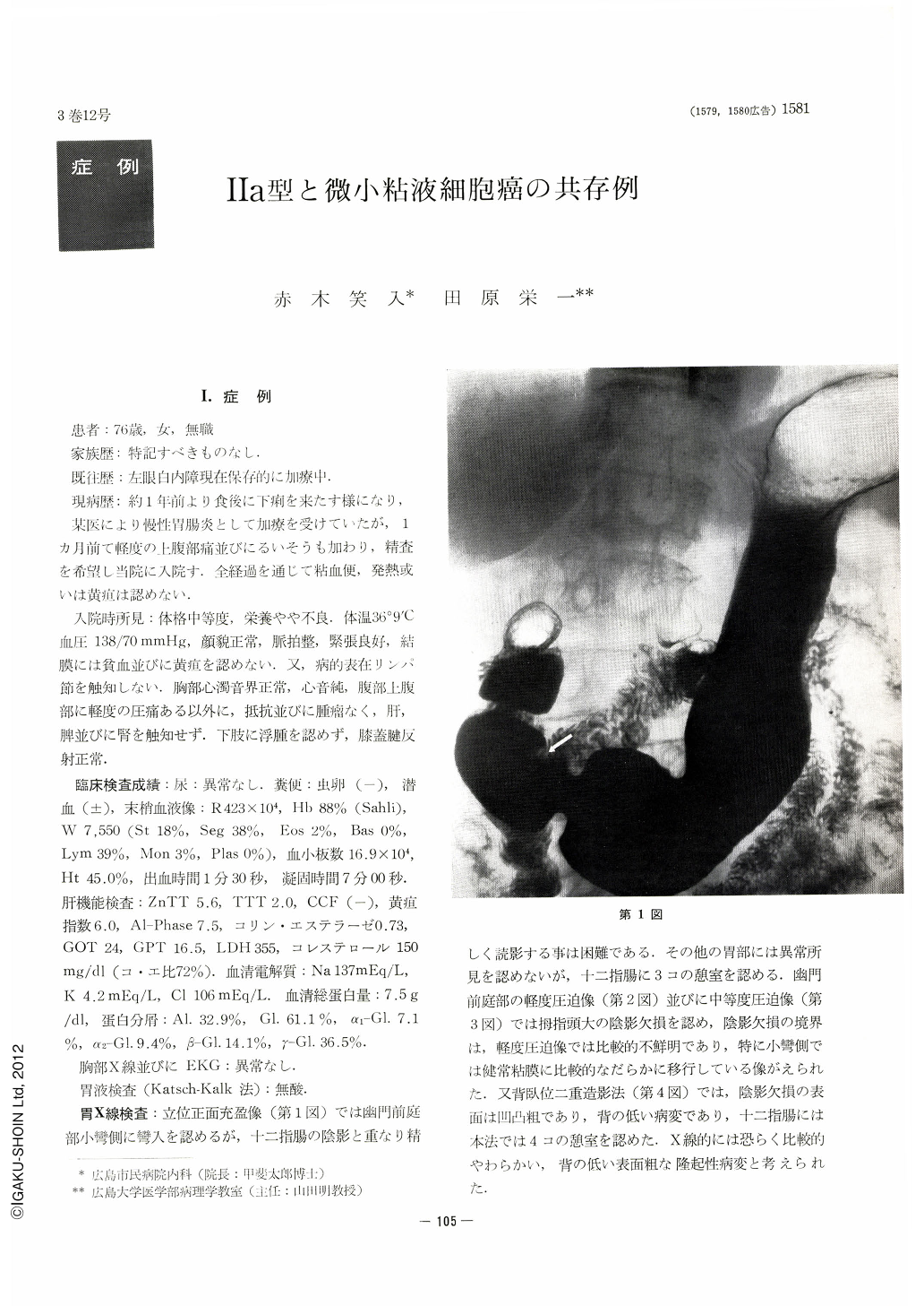
The x-ray examination (upright position, frontal view) in barium filled viscus revealed no particular disorder except slight concavity on the lesser curvature in the pyrolic antrum. Compression study brought out a filling defect the size of a thumb, the borders of which were comparatively indistinct, gradually melting into the normal mucous membrane on the lesser curvature side in particular. By double contrast technique, the surface of the image deficiency was visualized as uneven and coarse, suggestive of a relatively soft and slight protruding lesion with rough surface.
Through the use of endoscope a low and uneven protuberance was noticed on the posterior wall of the pyrolic antrum. It was sharply demarcated all around against surrounding mucosa, and the color of the surface was a bit faded. Though it was clearly irregular and uneven, no other surface peculiarities such as bleeding, rubescence or erosion were detected.
Macroscopical observation of the resected specimen revealed an uneven protrusion the size of 27×13 mm. As to the borders, these toward the cardiac orifice, the greater curve and the pylorus were clear. The border on the side of the lesser curvature was indistinct as it gradually merged into the surrounding mucous membrane. The surface of the elevation was coarse and uneven, resembling the unevenness of a flower-bed, a macroscopic clear sign of Ⅱa type early gastric cancer.
Histologically this Ⅱa type intraepithelial cancer proved to be papillary adenocarcinoma, atrophic hyperplastic gastritis probably forming the background of the process of its development. An amazing discovery was the exisitence of a very minute lesion lcm distal to the Ⅱa type early cancer, which naturally defied our combined examinations of x-ray and endoscopy and even macroscopical observation of the resected specimen as well. There was no change whatsoever on the surface of the mucous membrane, and the lesion was confined to single lumen of pyrolic glands, to be confirmed only by histological examination (mucoid carcinoma).
The coexistence of such a minute lesion with Ⅱa type intraepithelial cancer provides, in our opinion, an important key toward the problem of multicentric origin of cancer, the relation between intraepithelial cancer and hyperplastic gastritis, and furtheremore toward the elucidation of the origin and the proliferative process of mucoid carcinoma.

Copyright © 1968, Igaku-Shoin Ltd. All rights reserved.


