Japanese
English
- 有料閲覧
- Abstract 文献概要
- 1ページ目 Look Inside
はじめに
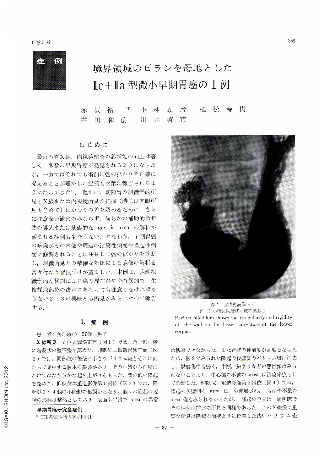
最近の胃X線,内視鏡検査の診断能の向上は著しく,多数の早期胃癌が発見されるようになったが,一方ではそれでも術前に癌の拡がりを正確に捉えることが難かしい症例も次第に報告されるようになってきた1).確かに,切除胃の組織学的所見とX線または内視鏡所見の把握(時には肉眼所見も含めて)にかなりの差を認めるために,さらに注意深い観察のみならず,何らかの補助的診断法の導入または基礎的なgastric areaの解析が望まれる症例も少なくない.すなわち,早期胃癌の病像がその内部や周辺の潰瘍性病変や隆起性病変に修飾されることに注目して癌の拡がりを診断し,組織所見との精確な対比による病像の解析を常々行なう習慣づけが望ましい.本例は,病理組織学的な検討による癌の局在がやや特異的で,生検採取部位の決定にあたっても注意しなければならない2,3の興味ある所見がみられたので報告する.
At both x-ray and endoscopic examinations of the stomach of a patient, a man 57 years of age, cancer was strongly suspected not only in a Ⅱc-like lesion extending around an erosion on the lesser curvature above the angle but also another nearby Ⅱc+Ⅱa-like lesion comprised of a cluster of small granular elevations. However, biopsy done twice proved negative Histological study revealed an early cancer, measuring 1.5 by 1.2 cm, a poorly differentiated adenocarcinoma with sm degree of depth invasion. These elevations as well as discolorations in the surrounding mucous membrane, found as they were in an area of multiple erosions, were thus presumed to be changes having developed in the course of repair of erosions. On that account, the pathological picture of these cancer lesions were variegated, with their intramural infiltration showing also unusual patterns. Of special interest is the fact that cancer lesion around the elevations was small, and within the protrusions themselves only their bases were invaded by cancer. Cancer partly encroached on the submucosal layer, but tops of the elevations were completely free from cancer. This is a case eloquently bespeaking the difliculty of preoperative diagnosis even including biopsy.

Copyright © 1971, Igaku-Shoin Ltd. All rights reserved.


