Japanese
English
- 有料閲覧
- Abstract 文献概要
- 1ページ目 Look Inside
はじめに
X線および内視鏡検査で多発性の潰瘍病変をともない,粘膜下腫瘍と鑑別困難であったⅡa+Ⅱc型早期胃癌の1症例を経験したので報告する.
A report is made of a case of IIa+IIc type early gastric cancer found in a 69-year-old female, very hard to distinguish from submucosal tumor by both x-ray and endoscopic examinations. It was besides associated with multiple ulcer lesions.
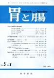
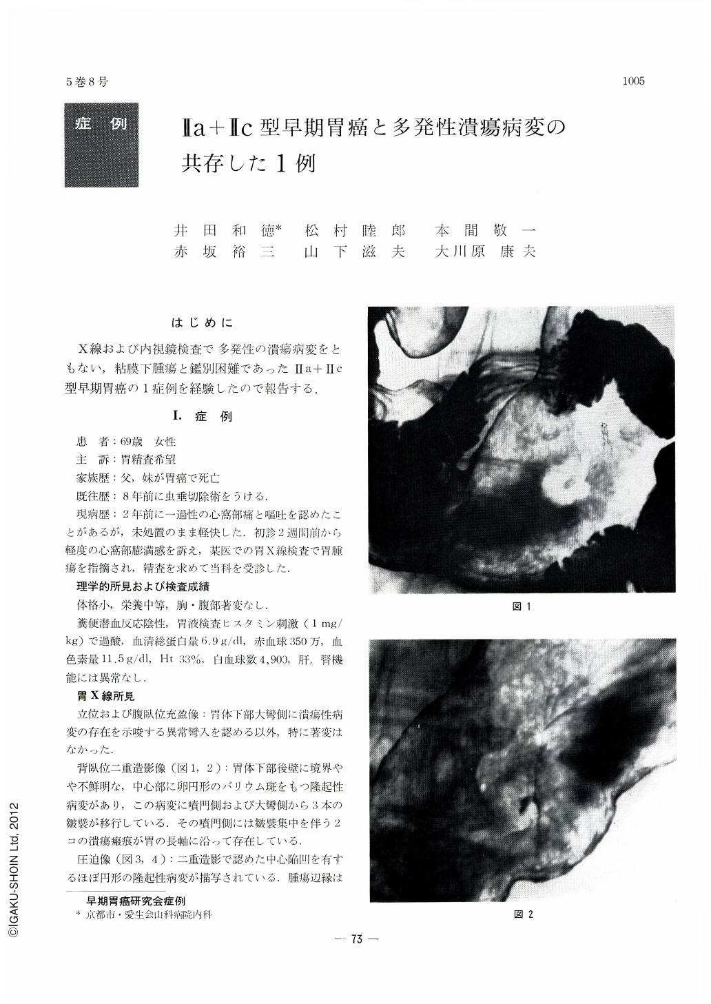
X-ray examination revealed a protruding lesion with uneven surface on the posterior wall of the lower corpus accompanied with a benign ulcer in the greater curvature side of it, in addition to 2 ulcer scars on its cardiac side. Although the protrusion was very suspicious of Ⅱa+Ⅱc type early cancer, it was impossible to rule out submucosal tumor because the margin of the tumor rose up relatively smoothly and several mucosal folds were recognized to pass into the lesion like bridging folds.
Endoscopy disclosed the mucosa around the protrusion gradually passing over into it without any change of appearance accompanied with mucosal folds as of bridging ones. Besides, neither engorgement nor erosion was recognized in the depression on the surface of the tumor, so that submucosal tumor was strongly suspected. Incidentally, the mucosal folds passing into the lesion were found to be continuous with ulcer lesions in the surrounding areas. Biopsy was the deciding factor in the diagnosis of the lesion, for cancer cells were demonstrated in the biopsied tissues taken from the depression on the surface of the protrusion.
The fresh specimen of the resected stomach showed a small protrusion, measuring 2.0 by 2.0cm, having a central depression. A shallow ulcer measuring 5mm in diameter was seen in the greater curvature side of the lesion in addition to ulcer scars on its cardiac side.
Histopathologically, it belonged to undifferentiated adenocarcinoma, its infiltration localized mostly within the submucosal layer. No cancer invasion was recognized on the mucosal surface.
This case is of interest not only because cancer infiltration looked like a submucosal tumor but also because the mucosal folds were modified in their patterns by the ulcer lesions in the surrounding areas as of bridging folds. Some comments on this case are further added.
Copyright © 1970, Igaku-Shoin Ltd. All rights reserved.