Japanese
English
- 有料閲覧
- Abstract 文献概要
- 1ページ目 Look Inside
はじめに
アニサキス幼虫による胃の好酸球性肉芽腫は決して珍らしいものではないが,その直上にⅡa型早期胃癌を合併した症例を経験した.
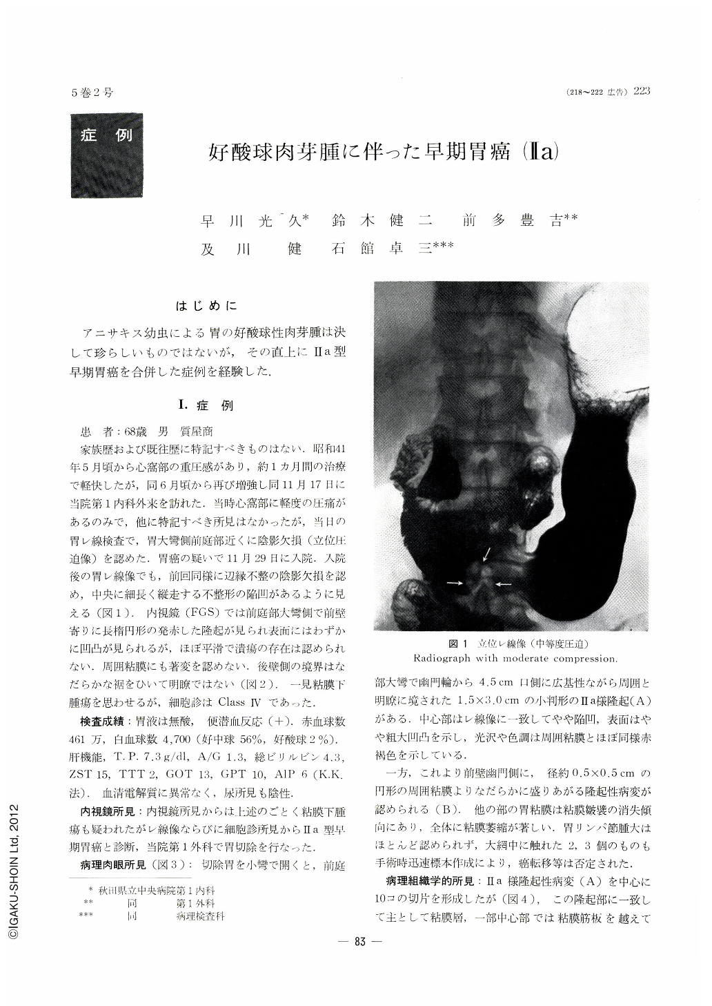
A 65-year-old man was admitted to the hospital because of epigastric dullness. Roentgenologically an irregular filling defect of uneven contour was demonstrated on moderate compression on the greater curvature of the antrum.
Fibergastroscopic picture revealed an elevated lesion on the anterior wall in the greater curvature side of the antrum with its surface slightly irregular. Cytologically it belonged to class Ⅳ.
Resectecl gross specimen presented two elevations in the antrum, the one (A) measuring 15×30 mm and the other (B) 5×5 mm. Histologically the former was adenocarcinoma tubulare (SAT Ⅱ, CAT 2) and the latter hyperplastic mucosal epithelia. Carcinomatous invasion of the lesion A was limited only to the submucosal layer, while histologically no malignancy was founcl on the elevation B.
In the microscopic preparation of the cross section 6 there was found an eosinophilic granuloma beneath the carcinomatous lesion A, and a worm body, partly destructed and deformed, was seen surrounded by infiltration of eosinophile cells. Radial and zonal arrangent of fibroblasts and surrounding fibrosis with infiltration of eosinophile cells suggest that the granuloma was formed by the invasion into the gastric mucosa of a larval “anisakis”. Concomitant early gastric cancer in this case is considered as merely incidental.

Copyright © 1970, Igaku-Shoin Ltd. All rights reserved.


