Japanese
English
- 有料閲覧
- Abstract 文献概要
- 1ページ目 Look Inside
- サイト内被引用 Cited by
Ⅰ.歴史的考察
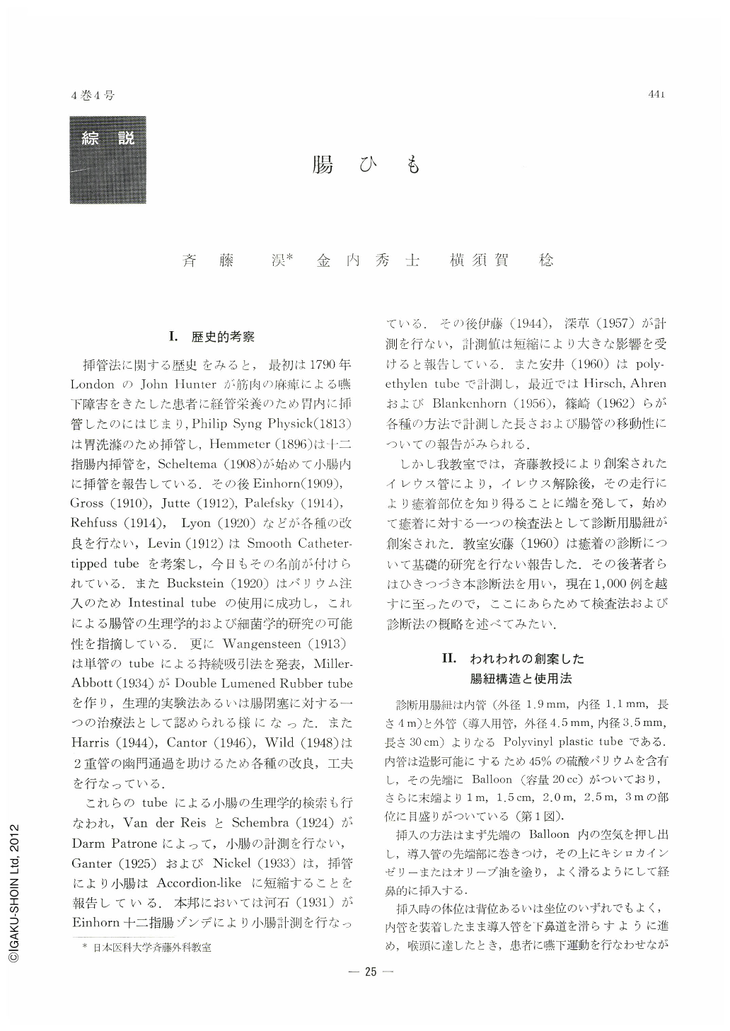
挿管法に関する歴史をみると,最初は1790年LondonのJohn Hunterが筋肉の麻痺による嚥下障害をきたした患者に経管栄養のため胃内に挿管したのにはじまり,Philip Syng Physick(1813)は胃洗源のため挿管し,Hemmeter(1896)は十二指腸内挿管を,Scheltema(1908)が始めて小腸内に挿管を報告している.その後Einhorn(1909),Gross(1910),Jutte(1912),Palefsky(1914),Rehfuss(1914),Lyon(1920)などが各種の改良を行ない,Levin(1912)はSmooth Catheter-tipped tubeを考案し,今日もその名前が付けられている.またBuckstein(1920)はバリウム注入のためIntestinal tubeの使用に成功し,これによる腸管の生理学的および細菌学的研究の可能性を指摘している.更にWangensteen(1913)は単管のtubeによる持続吸引法を発表,Miller-Abbott(1934)がDouble Lumened Rubber tubeを作り,生理的実験法あるいは腸閉塞に対する一つの治療法として認められる様になった.またHarris(1944),Cantor(1946),Wild(1948)は2重管の幽門通過を助けるため各種の改良,工夫を行なっている.
これらのtubeによる小腸の生理学的検索も行なわれ,Van der ReisとSchembra(1924)がDarm Patroneによって,小腸の計測を行ない,Ganter(1925)およびNickel(1933)は,挿管により小腸はAccordion-likeに短縮することを報告している.本邦においては河石(1931)がEinhorn十二指腸ゾンデにより小腸計測を行なっている.その後伊藤(1944),深草(1957)が計測を行ない,計測値は短縮により大きな影響を受けると報告している.また安井(1960)はpoly-ethylen tubeで計測し,最近ではHirsch,AhrenおよびBlankenhorn(1956),篠崎(1962)らが各種の方法で計測した長さおよび腸管の移動性についての報告がみられる.
Although intestinal adhesion is often diagnosed with ease, knowledge of its real state is as often limited. Examination by means of peroral barium meal, hithero extensively employed, is nevertheless insufiicient to locate exact region of adhesion, its length or its extent, a subject of great importance closely connected with surgical treatment. To supplement this insufilciency, the authors originated a diagnostic intestinal string, which is a 4 meters long radiopaque polyvinyl plastic string, consisting of two, outer and inner, tubes. The outer one, greater in diameter than the inner one, is used for smoothly introducing the inner tube down into the gullet. To the latter's dital tip is attached a small baloon, which is swallowed down, after having been deflated, along with the outer tube. When the tip of the baloon reaches the middle past of the esophagus, about 15 ml of water or gastrografin is then injected through the intestinal string into the baloon. A knot is made around the oral end of the inner tube to protect against reflux of water or contrast medium. After having been swallowed down with diet administered, the baloon as well as the inner tube will pass easily through the pyrolus down into the duodenum, being propelled distalwarcl out of the small intestine into the colon, soon to be excreted along with feces. Usually it takes about three days for the string to be discharged. X-ray pictures of patient with this string in his digestive organ are frequntly exposed so as to know exact condition of his adhesion. Sometimes a patient would undergo surgical intervention with his string remaining in his abdomen, when complicated adhesions exist. Abnormal run of the intestine and the colon in its various phases is thus checked by fluoroscopy.
By this diagnostic procedure of intestinal adhesion, accounting for more than 80 per cent in its accuracy, adhered intestine is classified in three types according to the alteration of the run of the bowel induced by traction of the string; (1) fixed type, (2) parallel type, and (3) similar type. They are explained as follows.
1. Fixed type. When a segment of the intestine is adhered to the abdominal wall, it remains fixed in spite of postural change.
2. Parallel type. When two remote intestinal segments are adhered, the distance between the two does not change by alteration of patient's posture.
3. Similar type. When two neighboring segments of the intestine are adhered to each other, the run of the adhered region of the bowel as visualized by the radiopaque string retains its original shape in spite of postural change.
On the basis of more than 1,000 cases of intestinal adhesion examined by this method in the authors' department, this procedure, not only of use to the diagnosis of intestinal adhesion, but to far wider diagnostic possibility as well, can be utilized for the time being in ascertaining; (1) the length of the intestine; (2) the site and the run of the intestine as well as its relation to other adjacent organs; (3) real phases of stagnation, reflux, and rotation of intestinal contents; (4) location of intestinal fistula or herniation and its relation to other organs; and (5) the site of anastomosis previous done, with no mistake of the run and direction of the bowel, when operation is done with the string remaining in his abdomen. Not only is this procedure expedient to performing surgical operation with safety and reliability, but also has proved to be of great service in elucidating the fact that various symptoms pertaining to adhesion or anastomosis do originate in the stagnation or abnormal rotation of intestinal contents.

Copyright © 1969, Igaku-Shoin Ltd. All rights reserved.


