Japanese
English
- 有料閲覧
- Abstract 文献概要
- 1ページ目 Look Inside
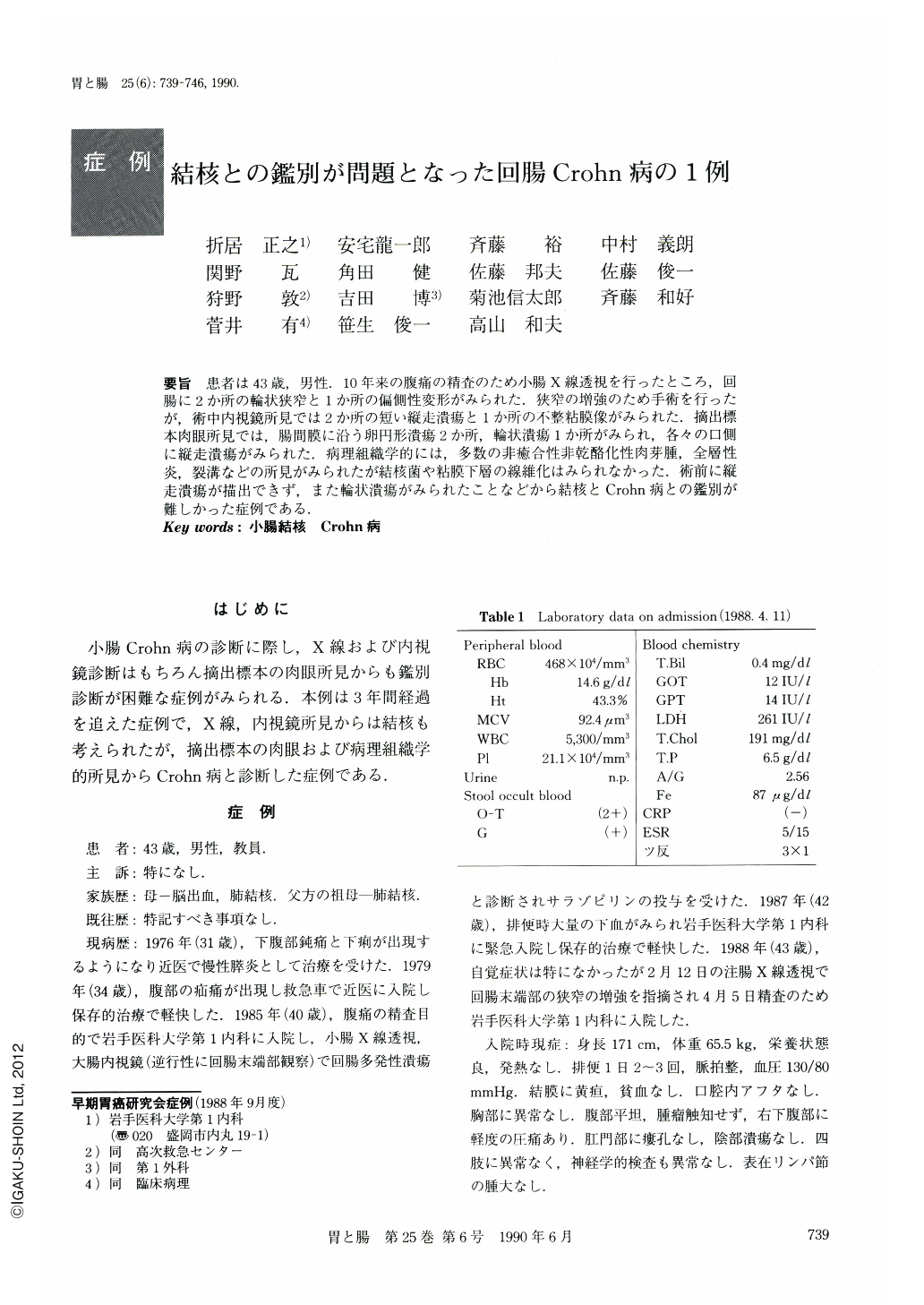
要旨 患者は43歳,男性.10年来の腹痛の精査のため小腸X線透視を行ったところ,回腸に2か所の輪状狭窄と1か所の偏側性変形がみられた.狭窄の増強のため手術を行ったが,術中内視鏡所見では2か所の短い縦走潰瘍と1か所の不整粘膜像がみられた.摘出標本肉眼所見では,腸間膜に沿う卵円形潰瘍2か所,輪状潰瘍1か所がみられ,各々の口側
に縦走潰瘍がみられた.病理組織学的には,多数の非癒合性非乾酪化性肉芽腫,全層性炎,裂溝などの所見がみられたが結核菌や粘膜下層の線維化はみられなかった.術前に縦走潰瘍が描出できず,また輪状潰瘍がみられたことなどから結核とCrohn病との鑑別が難しかった症例である.
We describe here a 43-year-old man with Crohn's disease very difficult to differentiate from tuberculosis.
At the age of 31 he started to have lower abdominal pain and diarrhea. Since 1985 melena and lower abdominal pain occurred so frequently that he was admitted to our hospital to undergo full investigation. His general condition was good, but slight tenderness was noticed in the right lower abdomen. Results of laboratory examination were unremarkable except for positive occult blood in the stool.
The double contrast study of the small intestine showed two areas of circular stenosis and an eccentric narrowing with pseudodiverticulum (Fig. 1 a, b). Endoscopic study revealed two short longitudinal ulcers with converging folds (lesion ①②) and irregular mucosa with irregular small ulcers (lesion ③) (Fig. 5 a-c).
The resected ileum showed an oval ulcer, irregular ulcer, circular shallow ulcer and longitudinal ulcer running along the mesenteric side (Fig. 6 a, b, c, e).
There were multiple non-caseating granulomas, transmural inflammation with aggregated pattern and fissuring ulcer, but a mucosal atrophy with ulcer scar was not seen (Fig. 7 a-f, 8).
X-ray study and endoscopic findings were compatible with the features of either intestinal tuberculosis or Crohn's disease. Subsequent macroscopic and histopathologic examinations showed the features of Crohn's disease which we believe was the definitive diagnosis.

Copyright © 1990, Igaku-Shoin Ltd. All rights reserved.


