Japanese
English
- 有料閲覧
- Abstract 文献概要
- 1ページ目 Look Inside
- サイト内被引用 Cited by
Ⅰ.はじめに
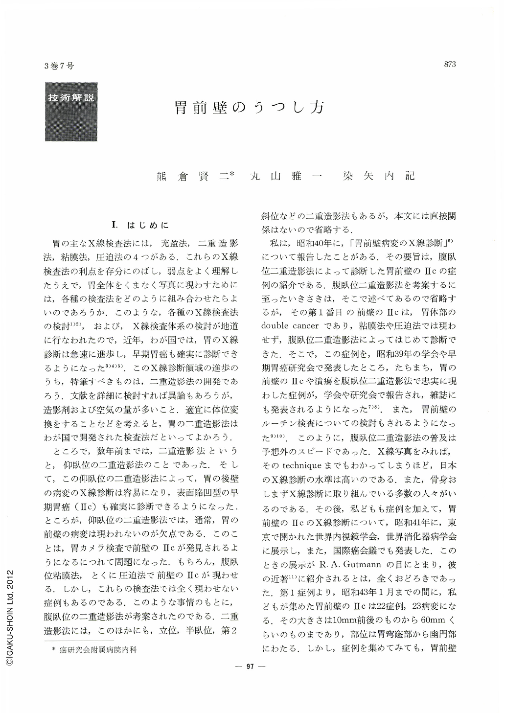
胃の主なX線検査法には,充盈法,二重造影法,粘膜法,圧迫法の4つがある.これらのX線検査法の利点を存分にのばし,弱点をよく理解したうえで,胃全体をくまなく写真に現わすためには,各種の検査法をどのように組み合わせたらよいのであろうか.このような,各種のX線検査法の検討1)2),および,X線検査体系の検討が地道に行なわれたので,近年,わが国では,胃のX線診断は急速に進歩し,早期胃癌も確実に診断できるようになった3)4)5).このX線診断領域の進歩のうち,特筆すべきものは,二重造影法の開発であろう.文献を詳細に検討すれば異論もあろうが,造影剤および空気の量が多いこと.適宜に体位変換をすることなどを考えると,胃の二重造影法はわが国で開発された検査法だといってよかろう.
ところで,数年前までは,二重造影法というと,仰臥位の二重造影法のことであった.そして,この仰臥位の二重造影法によって,胃の後壁の病変のX線診断は容易になり,表面陥凹型の早期胃癌(Ⅱc)も確実に診断できるようになった.ところが,仰臥位の二重造影法では,通常,胃の前壁の病変は現われないのが欠点である.このことは,胃カメラ検査で前壁のⅡcが発見されるようになるにつれて問題になった.もちろん,腹臥位粘膜法,とくに圧迫法で前壁のⅡcが現わせる.しかし,これらの検査法では全く現わせない症例もあるのである.このような事情のもとに,腹臥位の二重造影法が考案されたのである.二重造影法には,このほかにも,立位,半臥位,第2斜位などの二重造影法もあるが,本文には直接関係はないので省略する.
私は,昭和40年に,「胃前壁病変のX線診断」6)について報告したことがある.その要旨は,腹臥位二重造影法によって診断した胃前壁のⅡcの症例の紹介である.腹臥位二重造影法を考案するに至ったいきさきは,そこで述べてあるので省略するが,その第1番目の前壁のⅡcは,胃体部のdouble cancerであり,粘膜法や圧迫法では現わせず,腹臥位二重造影法によってはじめて診断できた.そこで,この症例を,昭和39年の学会や早期胃癌研究会で発表したところ,たちまち,胃の前壁のⅡcや潰瘍を腹臥位二重造影法で忠実に現わした症例が,学会や研究会で報告され,雑誌にも発表されるようになった7)8).また,胃前壁のルーチン検査についての検討もされるようになった9)10).このように,腹臥位二重造影法の普及は予想外のスピードであった.X線写真をみれば,そのtechniqueまでもわかってしまうほど,日本のX線診断の水準は高いのである.また,骨身おしまずX線診断に取り組んでいる多数の人々がいるのである.その後,私どもも症例を加えて,胃前壁のⅡcのX線診断について,昭和41年に,東京で開かれた世界内視鏡学会,世界消化器病学会に展示し,また,国際癌会議でも発表した.このときの展示がR. A. Gutmannの目にとまり,彼の近著11)に紹介されるとは,全くおどろきであった.第1症例より,昭和43年1月までの間に,私どもが集めた胃前壁のⅡcは22症例,23病変になる.その大きさは10mm前後のものから60mmくらいのものまであり,部位は胃穹窿部から幽門部にわたる.しかし,症例を集めてみても,胃前壁病変の写し方についての私どもの考え方は,昭和40年当時と余り変っていない.そのため,前の論文の繰り返しになるが,胃前壁病変の写し方について,私どもの集めたⅡcの症例にもとづき,私どもの考えを述べることにする.代表的な3症例を供覧する.
Although advanced cancer, large ulcer and polyp in the anterior wall of the stomach may be relatively easily diagnosed by the conventional x-ray examination, diagnostic difficulty arises in diagnosing minute lesions such as superficial depressed type of early cancer (Type Ⅱc).
Consideration was laid upon the method of x-ray demonstration of Type Ⅱc, based on the cases experienced until present. If ulcer scar is coexistent in cancerous as lesion observed in Type Ⅱc+Ⅲ, deformity of the stomch may be noted in the prone or upright barium-filled fillms, and if a part of Type Ⅱc rides on the lesser or greater curvature, marginal irregularity or depression may appear in them. But conclusive diagnosis can not bedrawn by the interpretation of those films.
Supine double contrast films may sometimes leave a print of Type Ⅱc in the anterior wall, but the conventionally employed method may be insufficient to diagnose the minute lesion in the anterior wall in contrast to the fact that Type Ⅱc in the anterior wall has been relatively easily diagnosed by the endoscopic examination.
To get an excellent detail of anterior lesion, compression study and prone double contrast method are indispensable. Compression study may be better, if a lesion is relatively small, but this method may fail to demonstrate large lesions.
In constidering such a circumstances the author emphasized the superiority of the prone double contrast method, and the role of the prone double contrast method and the effective sequence of each examination method in the routine x-ray work were discussed.
Three representative cases were displayed for the convenience of explanatory description on the prone double contrast method.

Copyright © 1968, Igaku-Shoin Ltd. All rights reserved.


