Japanese
English
- 有料閲覧
- Abstract 文献概要
- 1ページ目 Look Inside
症例
患者:内○初○,72歳,男
初診:昭和42年4月13日
手術:昭和42年8月18日
主訴:心窩部重圧感
既往歴:2~3年前より高血圧症として加療中である.又,若い頃より度々胃腸障害があったという.
家族歴:特記事項なし
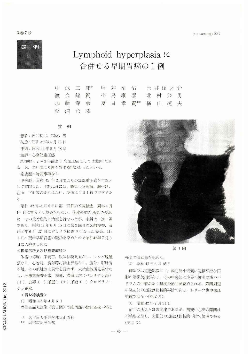
Patient was 72 years old male. February pressure feeling at epigastrium was arised. April ‘67’ first X-ray and gastro-camera examination were performed. Pathological change was situated at lesser curvature of pylorus. By gastro-camera finding, Ⅱa+Ⅱc type cancer was considered, but elevation around the margin was smooth and no color change was noticed, so this was tought to be erosion. Chief complaint hanged in the balance by symptomatic treatment. June and July '67, X-ray and gastro-camera examination were also repeated. By X-ray, shadow defect was noticed at the same part. Excavation was seen at the centre but surrounding elevation was smooth. By gastro-camera examination, reddening around white fur became prominent and the elevation around it became clear. Hard feeling seemed to be increased.
On July, elevation became hemisphere like tumor and another low elevation with smooth surface was also noticed at the oral side of the tumor. Diagnosis was made as Ⅱa+Ⅱc type early cancer but also possibility to be submucosal tumor was considered.
Operation was performed on August. Tumor, 20×17 mm in size, was seen at the lesser curvature of pylorus. Elevated change, 10×7 mm in size was noticed at the oral side of the tumor and considered as submucosal tumor.
Histologically, tumors were restricted inside of submucosal layer and consisted of multiplication of lymphatic tissue infiltrated with cancer cells. No cancer tissue was noticed outside of lymphatic tissue. Cancer was carcinoma adenotublare pathologically.

Copyright © 1968, Igaku-Shoin Ltd. All rights reserved.


