Japanese
English
- 有料閲覧
PPV購入案内
1,320円 (1,200円+税10%) こちらは、485ページから489ページの文献です。購入には医書.jp本会員登録が必要です。
会員登録完了後に改めて上記「購入サイトへ」を選択してください。
または「購入サイトへ」選択後に「購入ログイン」、「新規会員登録」の順に進んでください。
会員登録について詳しくはをご確認ください。
- Abstract 文献概要
- 1ページ目 Look Inside
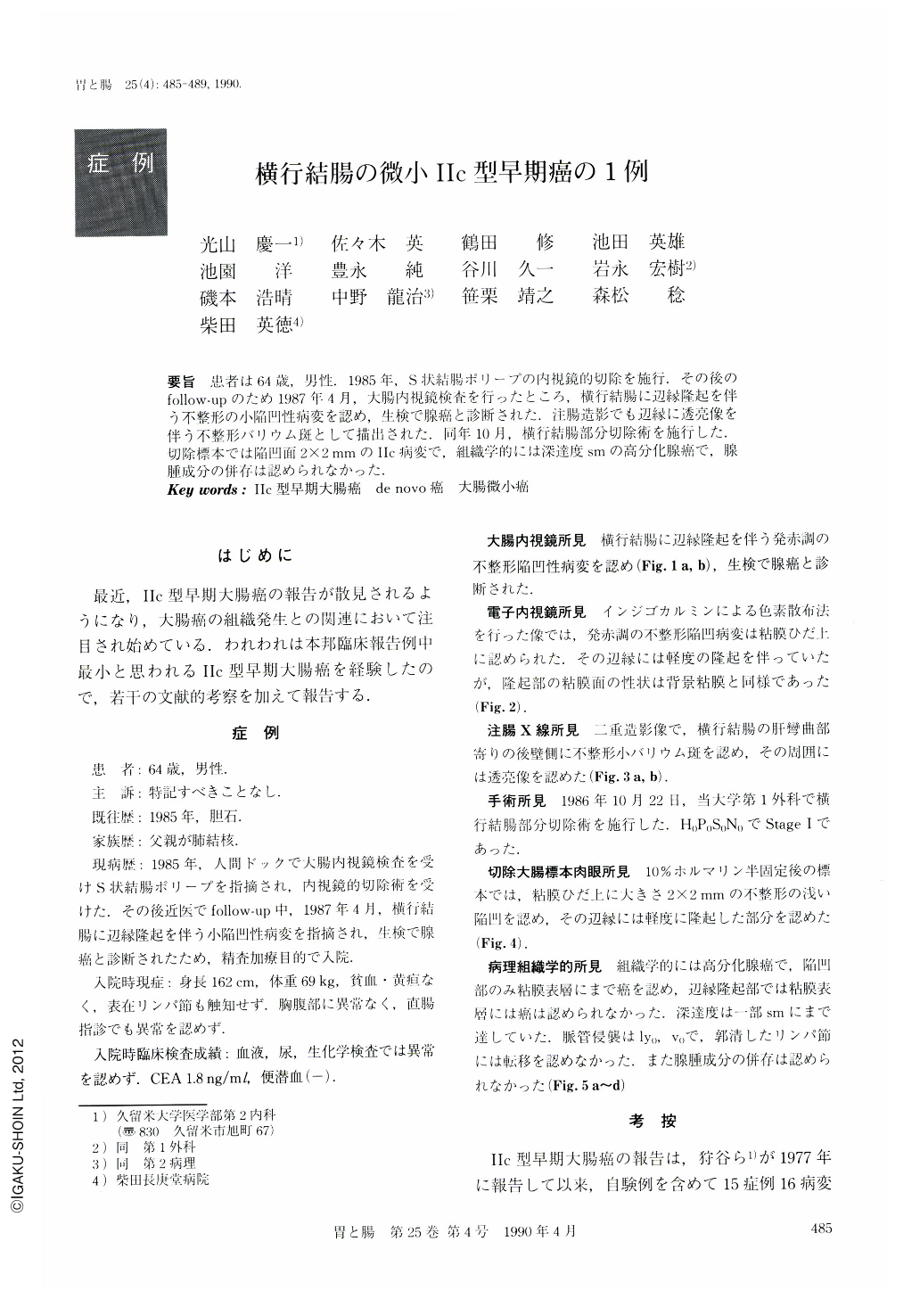
要旨 患者は64歳,男性.1985年,S状結腸ポリープの内視鏡的切除を施行.その後のfollow-upのため1987年4月,大腸内視鏡検査を行ったところ,横行結腸に辺縁隆起を伴う不整形の小陥凹性病変を認め,生検で腺癌と診断された.注腸造影でも辺縁に透亮像を伴う不整形バリウム斑として描出された.同年10月,横行結腸部分切除術を施行した.切除標本では陥凹面2×2mmのⅡc病変で,組織学的には深達度smの高分化腺癌で,腺腫成分の併存は認められなかった.
A 64-year-old man was addmitted to Kurume University Hospital for further medical evaluation and treatment of a minute depressed lesion in the transverse colon.
He had previously undergone endoscopic polypectomy of sigmoid polyps in 1985. Follow-up endoscopy in April 1987 disclosed a small, reddish and irregular depressed lesion with surrounding elevation in the transverse colon. Biopsy specimen showed adenocarcinoma.
Partial resection of the transverse colon was performed on October 22, 1987. Surgical specimen showed a minute and shallow depressed lesion, measuring 2 × 2 mm in size. The lesion was histologically diagnosed as well differentiated adenocarcinoma with submucosal invasion. No adenomatous component was contained. Finally, the lesion was proven to be Ⅱc type "de novo" cancer of the colon.
Copyright © 1990, Igaku-Shoin Ltd. All rights reserved.