Japanese
English
- 有料閲覧
- Abstract 文献概要
- 1ページ目 Look Inside
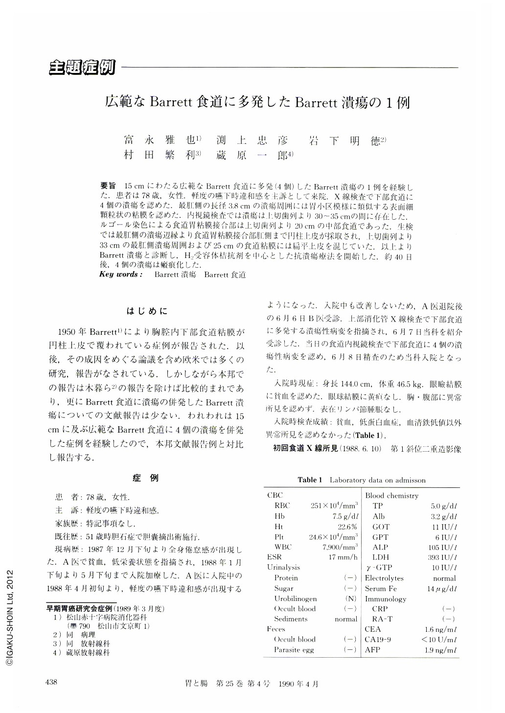
要旨 15cmにわたる広範なBarrett食道に多発(4個)したBarrett潰瘍の1例を経験した.患者は78歳,女性.軽度の嚥下時違和感を主訴として来院.X線検査で下部食道に4個の潰瘍を認めた.最肛側の長径3.8cmの潰瘍周囲には胃小区模様に類似する表面細顆粒状の粘膜を認めた.内視鏡検査では潰瘍は上切歯列より30~35cmの間に存在した.ルゴール染色による食道胃粘膜接合部は上切歯列より20cmの中部食道であった.生検では最肛側の潰瘍辺縁より食道胃粘膜接合部肛側まで円柱上皮が採取され,上切歯列より33cmの最肛側潰瘍周囲および25cmの食道粘膜には扁平上皮を混じていた.以上よりBarrett潰瘍と診断し,H2受容体桔抗剤を中心とした抗潰瘍療法を開始した.約40日後,4個の潰瘍は瘢痕化した.
A 78-year-old woman was admitted to our hospital with a complaint of mild dysphagia. Radiologic examination revealed four deep ulcers in the lower esophagus. The largest, approximately 3.8 cm in size, and most distally located ulcer was surrounded by fine granular mucosa resembling gastric mucosa. Esophagoscopy revealed four ulcers at the level of 30-35 cm from the incisors. The squamo-columnar junction was clearly determined in the middle esophagus (20 cm from the incisors) by Lugol-staining. Multiple biopsy specimens obtained from the ulcers and the squamo-columnar junction revealed columnar epithelium as well as squamous epithelium in part. The diagnosis of Barrett's ulcer was thus made. Treatment was commenced with antipeptic ulcer drugs including Famotidine 60 mg/day. The ulcers healed in about 40 days.

Copyright © 1990, Igaku-Shoin Ltd. All rights reserved.


