Japanese
English
- 有料閲覧
- Abstract 文献概要
- 1ページ目 Look Inside
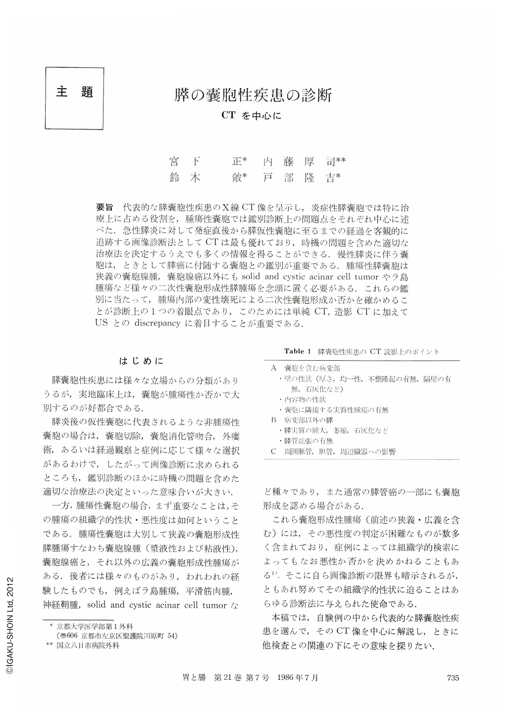
要旨 代表的な膵囊胞性疾患のX線CT像を呈示し,炎症性膵囊胞では特に治療上に占める役割を,腫瘍性囊胞では鑑別診断上の問題点をそれぞれ中心に述べた.急性膵炎に対して発症直後から膵仮性囊胞に至るまでの経過を客観的に追跡する画像診断法としてCTは最も優れており,時機の問題を含めた適切な治療法を決定するうえでも多くの情報を得ることができる.慢性膵炎に伴う囊胞は,ときとして膵癌に付随する囊胞との鑑別が重要である.腫瘍性膵囊胞は狭義の囊胞腺腫,囊胞腺癌以外にもsolid and cystic acinar cell tumorやラ島腫瘍など様々の二次性囊胞形成性膵腫瘍を念頭に置く必要がある.これらの鑑別に当たって,腫瘍内部の変性壊死による二次性囊胞形成か否かを確かめることが診断上の1つの着眼点であり,このためには単純CT,造影CTに加えてUSとのdiscrepancyに着目することが重要である.
CT findings of various cystic lesions of the pancreas were demonstrated. CT is the best diagnostic tool for visualization of the changing image of the pancreas from the onset of acute pancreatitis to the maturation of pseudocyst. It also gives information helpful for the timing of surgical intervention, its method and approach. Pancreatic cancer featured by cyst forma-tion is sometimes misdiagnosed as only chronic pancreatitis with pseudocyst or retention cyst. Cystic neoplasms of the pancreas, in a broad sense, are comprised of various pancreatic tumors, cystadenocarcinoma, cystadenoma, solid and cystic acinar cell tumor, islet cell tumor with cystic degeneration and others. It is one of the diagnostic clues to discern whether the lesion is the primary cyst or the secondary one formed by degenerative change of solid neoplasm. For this purpose, the discrepancy between CT findings and US findings should be paid attention to.

Copyright © 1986, Igaku-Shoin Ltd. All rights reserved.


