Japanese
English
- 有料閲覧
- Abstract 文献概要
- 1ページ目 Look Inside
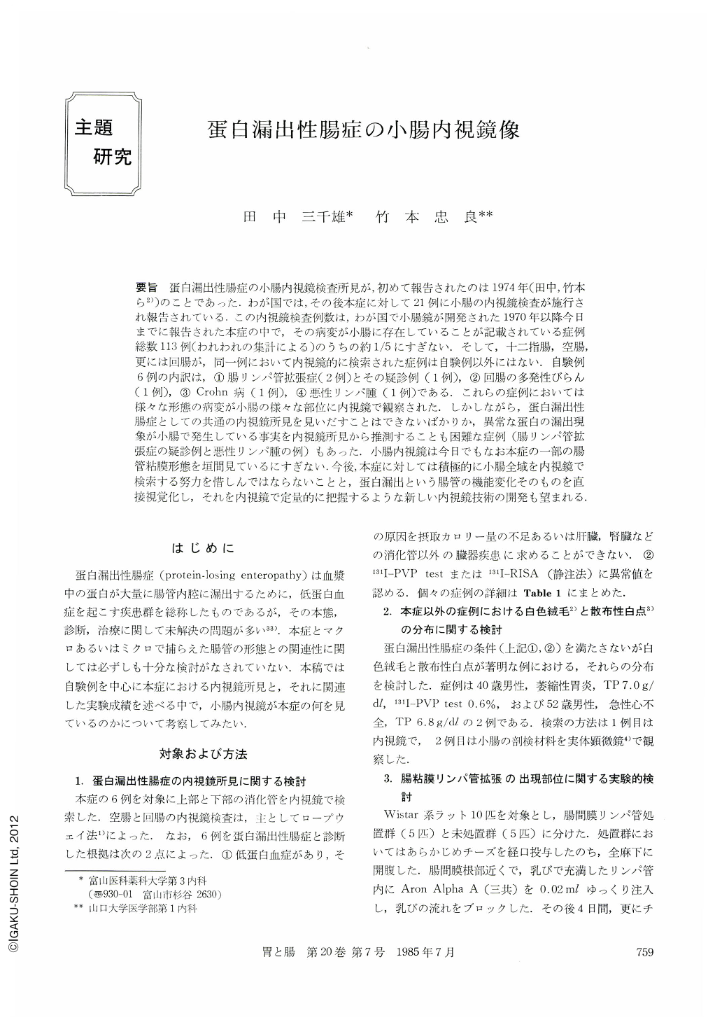
要旨 蛋白漏出性腸症の小腸内視鏡検査所見が,初めて報告されたのは1974年(田中,竹本ら2))のことであった.わが国では,その後本症に対して21例に小腸の内視鏡検査が施行され報告されている.この内視鏡検査例数は,わが国で小腸鏡が開発された1970年以降今日までに報告された本症の中で,その病変が小腸に存在していることが記載されている症例総数113例(われわれの集計による)のうちの約1/5にすぎない.そして,十二指腸,空腸,更には回腸が,同一例において内視鏡的に検索された症例は自験例以外にはない.自験例6例の内訳は,①腸リンパ管拡張症(2例)とその疑診例(1例),②回腸の多発性びらん(1例),③Crohn病(1例),④悪性リンパ腫(1例)である.これらの症例においては様々な形態の病変が小腸の様々な部位に内視鏡で観察された.しかしながら,蛋白漏出性腸症としての共通の内視鏡所見を見いだすことはできないばかりか,異常な蛋白の漏出現象が小腸で発生している事実を内視鏡所見から推測することも困難な症例(腸リンパ管拡張症の疑診例と悪性リンパ腫の例)もあった.小腸内視鏡は今日でもなお本症の一部の腸管粘膜形態を垣間見ているにすぎない.今後,本症に対しては積極的に小腸全域を内視鏡で検索する努力を惜しんではならないことと,蛋白漏出という腸管の機能変化そのものを直接視覚化し,それを内視鏡で定量的に把握するような新しい内視鏡技術の開発も望まれる.
Protein-losing enteropathy is a rather rare abnormal state of the intestinal tract. In Japan, 125 cases with protein-losing enteropathy have been reported in the past 15 years. And small intestinal diseases were found in 118 cases out of 125. The most frequent small intestinal disease combined with proteinlosing enteropathy was intestinal lymphangiectasia (63 cases).
Endoscopic findings of intestinal lymphangiectasia were first reported by us in 1974 (Case 1 in Table 1) 1).“Scattered white spot”, “white villus”(Fig. 1a) and color change at the villous tip (Fig. 1b) were noticed in the duodenal mucosa. Oozing of the chyle-like fluid was also observed after rubbing the mucosal surface endoscopically (Fig. 1c). Such findings were not recognized in the jejunum (Fig. 1d) and ileum. Dissecting microscopic and histological examination of biopsy specimens obtained from the duodenum was done. Partial defects of the villous tip were noticed under the dissecting microscope (Fig. 2a). This shape was very similar to that of villi observed by endoscopy (Fig. 1b). Scanning electron micrography revealed that the epithelial layer of the villus tip was exfoliated and the lamina propria mucosae was exposed (Fig. 3a and b). Histological studies including transmission electron microscopic examination revealed dilated central lacteal, extruded epithelial layer at villous tip (Fig. 4a), lipid droplets in the absorptive epithelial cell and intracellular space (Fig. 4b), dilatation of intracellular space, closure of tight junction (Fig. 5a) and protein-like substance and chylomicron-like substance in the dilated intracellular space (Fig. 5b).
After that first case report, we carried out endoscopic examination of the duodenum, jejunum and ileum in five cases with protein-losing enteropathy (Case 2~6 in Table 1). These five cases had the small intestinal diseases of lymphangiectasia (Fig. 6a~d), ileal erosions (Fig. 7a), Crohn's disease, malignant lymphoma (Fig. 7b and c) or thoracic duct occlusion with duodenal elevated lesion (Fig. 7d). It was very difficult to presume the relationship between endoscopic findings and the phenomenon of proteinlosing. For instance, in some cases without proteinlosing enteropathy,“scattered white spot”and“white villus”in the small intestine were as widely distributed as in cases with protein-losing enteropathy combined with intestinal lymphangiectasia (Fig. 8).
In Japan, small intestinal endoscopic findings have been reported in 26 cases with protein-losing enteropathy (Table 2). But most of these cases have been examined endoscopically within the limits to the duodenum and upper part of the jejunum.
We have succeeded in making“scattered white spot”by means of experimental blockage of rat's mesenteric lymph vessel (Fig. 10). Marked dilated villous lacteal filled with chylomicron was the characteristic of the histologic appearance of “scattered white spot”. The distribution of “scattered white spot” in rats (Fig. 9) suggests that endoscopic examination of intestinal lymphangiectasia should be done in all parts of the small intestine.
In summary, we reported endoscopic findings of six cases with protein-losing enteropathy in this paper. It was not easy to presume the relationship between endoscopic findings and the phenomenon of protein-losing. We expect that endoscopic examination will play an important role in the study on protein-losing enteropathy.

Copyright © 1985, Igaku-Shoin Ltd. All rights reserved.


