Japanese
English
- 有料閲覧
- Abstract 文献概要
- 1ページ目 Look Inside
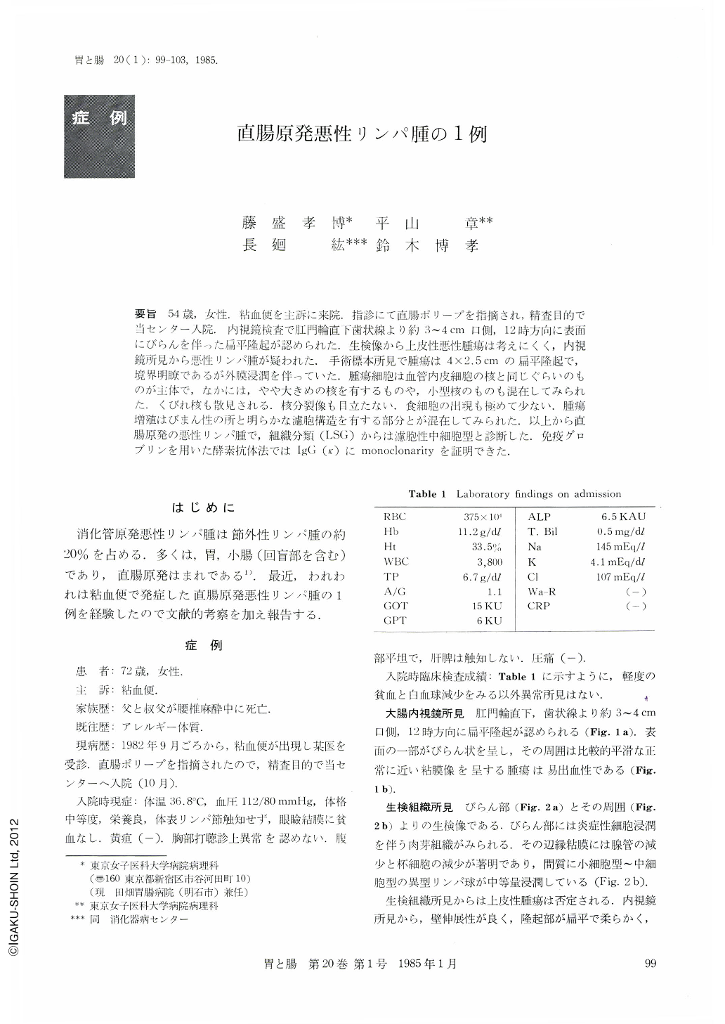
要旨 54歳,女性.粘血便を主訴に来院.指診にて直腸ポリープを指摘され,精査目的で当センター入院.内視鏡検査で肛門輪直下歯状線より約3~4cm口側,12時方向に表面にびらんを伴った扁平隆起が認められた.生検像から上皮性悪性腫瘍は考えにくく,内視鏡所見から悪性リンパ腫が疑われた.手術標本所見で腫瘍は4×2.5cmの扁平隆起で,境界明瞭であるが外膜浸潤を伴っていた.腫瘍細胞は血管内皮細胞の核と同じぐらいのものが主体で,なかには,やや大きめの核を有するものや,小型核のものも混在してみられた.くびれ核も散見される.核分裂像も目立たない.食細胞の出現も極めて少ない.腫瘍増殖はびまん性の所と明らかな濾胞構造を有する部分とが混在してみられた.以上から直腸原発の悪性リンパ腫で,組織分類(LSG)からは濾胞性中細胞型と診断した.免疫グロブリンを用いた酵素抗体法ではIgG(κ)にmonoclonarityを証明できた.
A 54 year-old woman patient visited our hospital with a chief complaint of bloody stool. Digital examination revealed a rectal mass. Colonoscopy showed a flat polypoid lesion with superficial erosion and minimal friability. Proctosigmoidectomy was done by the reason that its endoscopic finding was suggestive of malignant lymphoma, although the biopsy specimen revealed no tumor cells.
The tumor was limited in the rectum without any metastasis in the liver, spleen and retroperitoneal cavity. In the resected specimen the lesion, 40×25 mm in diameter, was located in the rectum, about 40 mm from the dentate line. In histologic examination, the most distinctive feature of tumor cells was the nodular pattern of growth at low-power view. Tumor cells were composed of small and large lymphoid cells (the population of large cells was less than 20%). Starry-sky cells were absent. Small to medium-sized cells with irregular nuclei showed occasional cleavage lines. The chromatin was coarse and the nucleoli were inconspicuous. Sometimes small plasma cells or plasmacytoid cells were observed in or among neoplastic cells. Immunoperoxidase studies demonstrated the monoclonal B-cell origin of these cells (lgG (κ)). By these findings, this tumor was diagnosed as primary rectal malignant lymphoma, follicular, mediumsized cell type (Classification of Japan Lymphoma-Leukemia Study Group; LSG), Stage Ⅰ.

Copyright © 1985, Igaku-Shoin Ltd. All rights reserved.


