Japanese
English
- 有料閲覧
- Abstract 文献概要
- 1ページ目 Look Inside
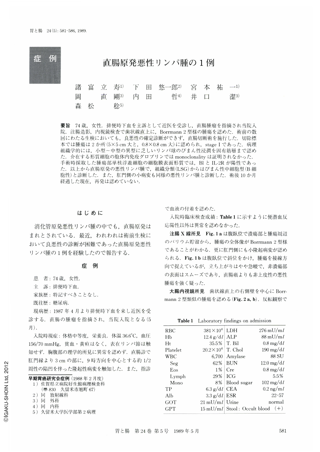
要旨 74歳,女性.排便時下血を主訴として近医を受診し,直腸腫瘤を指摘され当院入院.注腸造影,内視鏡検査で歯状線直上に,Borrmann 2型様の腫瘍を認めた.術前の数回にわたる生検においても,良悪性の確定診断ができず,直腸切断術を施行した.切除標本では腫瘍は2か所(5×5cm大と,0.8×0.8cm大)に認められ,stageⅠであった.病理組織学的には,小型~中型の異型に乏しいリンパ球のびまん性浸潤を固有筋層まで認めた.介在する形質細胞の胞体内免疫グロブリンではmonoclonalityは証明されなかった.手術時採取した腫瘍部単核浮遊細胞の細胞膜表面形質では,BlとIL-2Rが陽性であった.以上から直腸原発の悪性リンパ腫で,組織分類(LSG)からはびまん性中細胞型(B細胞性)と診断した.また,肛門側の小病変も同様の悪性リンパ腫と診断した.術後10か月経過した現在,再発は認めていない.
A 74-year-old woman visited our hospital complaning of bloody stool. Digital examination revealed a rectal mass. Irrigoscopy and colonoscopy showed a Borrmann 2-like lesion (Figs. 1a, and 2a). The biopsy specimens contained no atypical cells. Malignant lymphoma, however was suspected and Miles' operation was performed. Two stage Ⅰ lesions, 50×50 and 8×8 mm in di-ameter, respectively, were resected (Fig. 3). Upon histologic examination, the tumor cells, medium sized with no atypia, were distributed diffusely (Fig. 6). The tumor cells had invaded into the propria muscularis (Fig. 4). No monoclonality was observed in plasma cell immunoglobulin. Surface phenotype studies of the resected tumor cells were positive for IL-2 receptor and B-1 (Table 2). Based on these findings, these tumors were diagnosed as primary rectal malignant lymphoma diffuse, medium-sized cell type and B cell type (Classification of Japan Lymphoma―Leukemia Study Group : LSG). The patient is alive and well 10 months after surgery.

Copyright © 1989, Igaku-Shoin Ltd. All rights reserved.


