Japanese
English
- 有料閲覧
- Abstract 文献概要
- 1ページ目 Look Inside
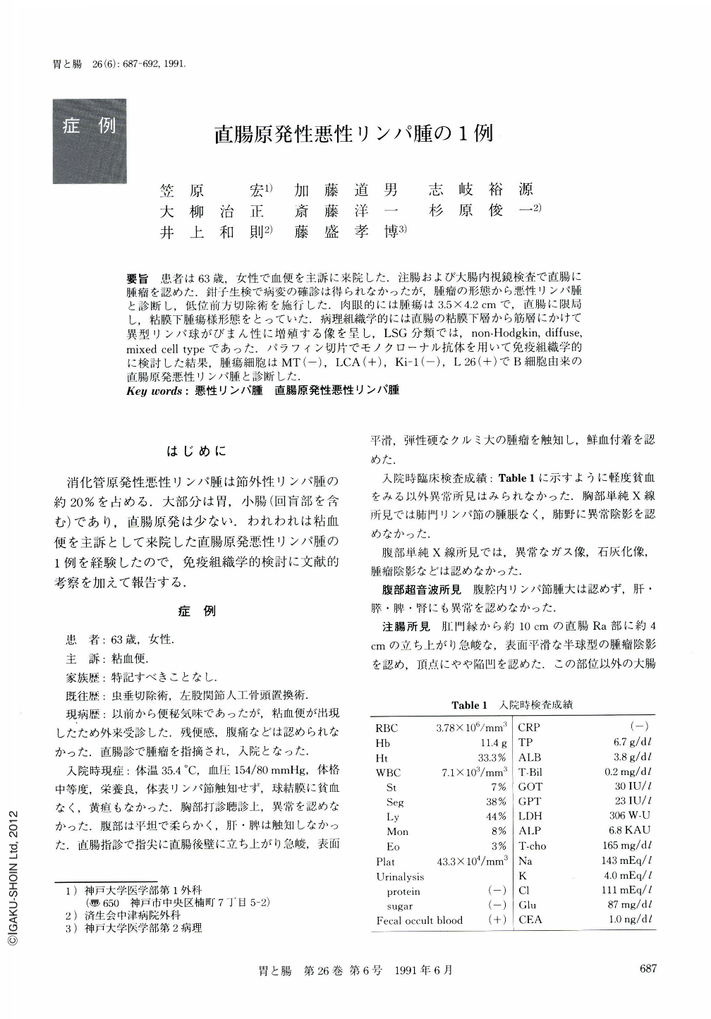
要旨 患者は63歳,女性で血便を主訴に来院した.注腸および大腸内視鏡検査で直腸に腫瘤を認めた.鉗子生検で病変の確診は得られなかったが,腫瘤の形態から悪性リンパ腫と診断し,低位前方切除術を施行した.肉眼的には腫瘍は3.5×4.2cmで,直腸に限局し,粘膜下腫瘍様形態をとっていた.病理組織学的には直腸の粘膜下層から筋層にかけて異型リンパ球がびまん性に増殖する像を呈し,LSG分類では,non-Hodgkin,diffuse,mixed cell typeであった.パラフィン切片でモノクローナル抗体を用いて免疫組織学的に検討した結果,腫瘍細胞はMT(-),LCA(+),Ki-1(-),L26(+)でB細胞由来の直腸原発悪性リンパ腫と診断した.
A 63 year-old woman patient was admitted with bloody stool as her chief complaint. Digital examination revealed a rectal mass. Barium enema (Fig. 2) and colonoscopy (Fig. 3) revealed a tumor of the rectum. Its endoscopic findings were suggestive of malignant lymphoma, although the biopsy specimen revealed no tumor cells. Under a clinical diagnosis of a malignant lymphoma of the rectum, a sphincter-saving operation with end-to-end anastomosis was performed. The tumor was limited to the rectum without any metastasis in the liver, spleen and retroperitoneal cavity. In the resected specimen the lesion, 3.5×1.2 cm in diameter, was located in the rectum (Fig. 4). In histologic examination, the most distinctive feature of the tumor cells was the diffuse pattern of growth at low-power view (Fig. 5 a). Tumor cells were composed of small lymphoid cells. Mitoses were found occasionally at high-power view (Fig. 5 b). Immunoperoxidase studies demonstrated the monoclonal B cell origin of these cells (LCA(+), L 26(+), MT 1(-), Ki-1(-)) (Fig. 6).
Using these findings, this tumor was diagnosed as primary rectal malignant lymphoma, diffuse mixed-cell type (classification of Japan Lymphoma-Leukemia Study Group; LSG), stage Ⅰ.

Copyright © 1991, Igaku-Shoin Ltd. All rights reserved.


