Japanese
English
- 有料閲覧
- Abstract 文献概要
- 1ページ目 Look Inside
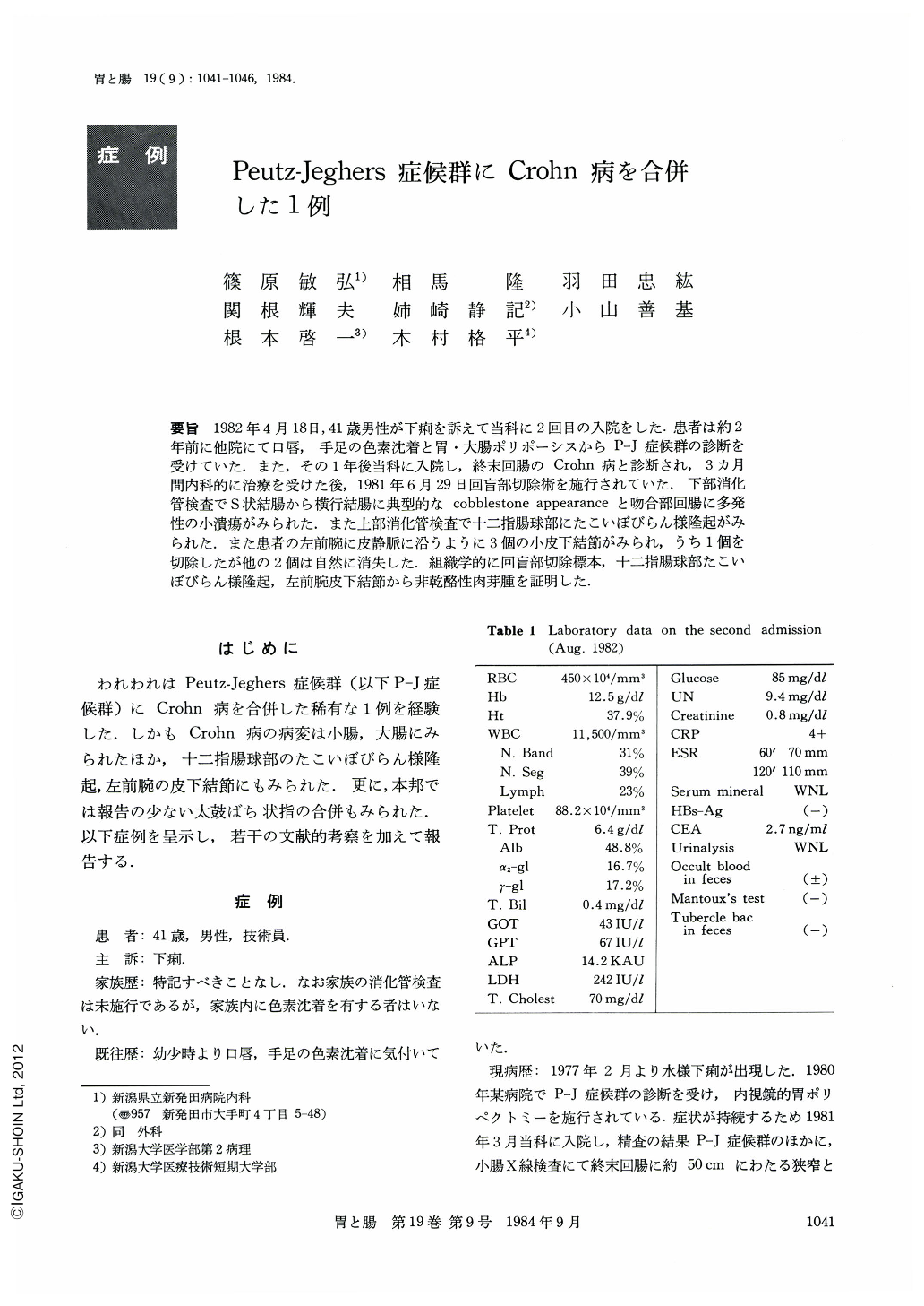
要旨 1982年4月18日,41歳男性が下痢を訴えて当科に2回目の入院をした.患者は約2年前に他院にて口唇,手足の色素沈着と胃・大腸ポリポーシスからP-J症候群の診断を受けていた.また,その1年後当科に入院し,終末回腸のCrohn病と診断され,3カ月間内科的に治療を受けた後,1981年6月29日回盲部切除術を施行されていた.下部消化管検査でS状結腸から横行結腸に典型的なcobble stoneappearanceと吻合部回腸に多発性の小潰瘍がみられた.また上部消化管検査で十二指腸球部にたこいぼびらん様隆起がみられた.また患者の左前腕に皮静脈に沿うように3個の小皮下結節がみられ,うち1個を切除したが他の2個は自然に消失した.組織学的に回盲部切除標本,十二指腸球部たこいぼびらん様隆起,左前腕皮下結節から非乾酪性肉芽腫を証明した.
A 41 year-old man was admitted to our hospital on August 18, 1982, complaining of diarrhea.
About two years ago, he was admitted to another hospital and was diagnosed as having Peutz-Jeghers syndrome because of gastric and colon polyposis with melanin pigmentation on his lips, hands and feet. One year after the first admission he was readmitted to our hospital and was diagnosed as having Crohn's disease of the terminal ileum. Conservative treatment for three months was done followed by a ileocecal resection in June 29, 1981.
Physical examination revealed melanin pigmentation and clubbed fingers. Radiologic and endoscopic examinations of lower intestine revealed typical cobblestone appearance from the upper sigmoid colon to the transverse colon and multiple small ulcers in the anastomotic ileum. And two polyps were shown in the colon and they were resected endoscopically. Upper gastrointestinal examinations showed erosive polypoid lesions in the duodenal bulb. Three small subcutaneous nodules were observed along the cutaneous vein in his left forearm. One of them was resected; thereafter the others disappeared spontaneously. By the histological analysis noncaseating epithelioid cell glanulomas were shown in the resected specimen of the ileum, and in an erosive polypoid lesion in the duodenal bulb as well as in a subcutaneous nodule.
The patient was treated with salicylazosulfapyridine and corticosteroid, and remission occurred after one month.

Copyright © 1984, Igaku-Shoin Ltd. All rights reserved.


