Japanese
English
- 有料閲覧
- Abstract 文献概要
- 1ページ目 Look Inside
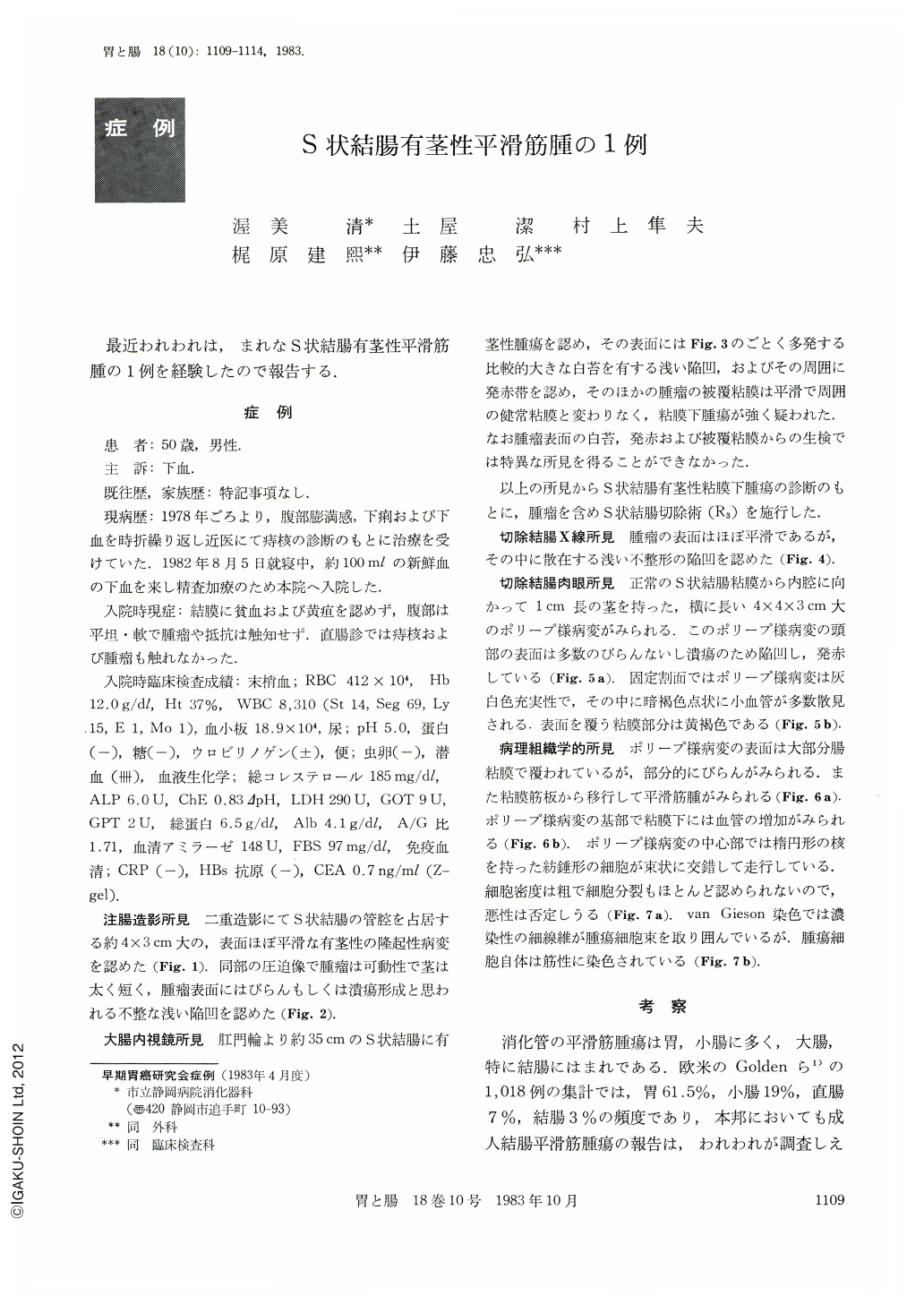
最近われわれは,まれなS状結腸有茎性平滑筋腫の1例を経験したので報告する.
症 例
患 者:50歳,男性.
主 訴:下血.
既往歴,家族歴:特記事項なし.
現病歴:1978年ごろより,腹部膨満感,下痢および下血を時折繰り返し近医にて痔核の診断のもとに治療を受けていた.1982年8月5日就寝中,約100mlの新鮮血の下血を来し精査加療のため本院へ入院した.
The patient, a man aged 54, was admitted to the hospital because of about 100ml of fresh anal bleeding while asleep. Laboratory data at admission were normal except for highly positive occult blood of stools. Barium enema examination revealed a pedunculated tumor in the sigmoid. Its surface showed irregular, shallow depression suggesting either erosion or ulceration. Colonofiberscopy showed a pedunculated tumor in the sigmoid about 35 cm oral from the anal ring. Its surface was spotted with multiple, relatively large shallow depressions with white coats surrounded by areas of redness. The remaining mucosa over the tumor was smooth the same as the normal mucosa. It was highly suggestive of a submucosal tumor. Biopsy gave us no definite clue to an accurate diagnosis. Therefore, under a diagnosis of a pedunculated submucosal tumor of the sigmoid colon its resection was carried out (R3).
Macroscopically, the surgical specimen showed a polypoid lesion, 4×4×3 cm, intraluminally protruding with a 1 cm long stalk. The surface of the tip was depressed and reddened because of a number of erosions or ulcers. The cut surface of the tumor was mostly covered with intestinal mucosa dotted with erosions and ulcers. The inner part was grey white and solid. Histologically, the tumor was filled with spindle-shaped cells with oval nuclei running criss-cross in bundles. Mitosis was hardly recognized, and the tumor was considered as a benign leiomyoma.
A reference was made to the literature of leiomyoma of colon in Japan.
Copyright © 1983, Igaku-Shoin Ltd. All rights reserved.