Japanese
English
- 有料閲覧
- Abstract 文献概要
- 1ページ目 Look Inside
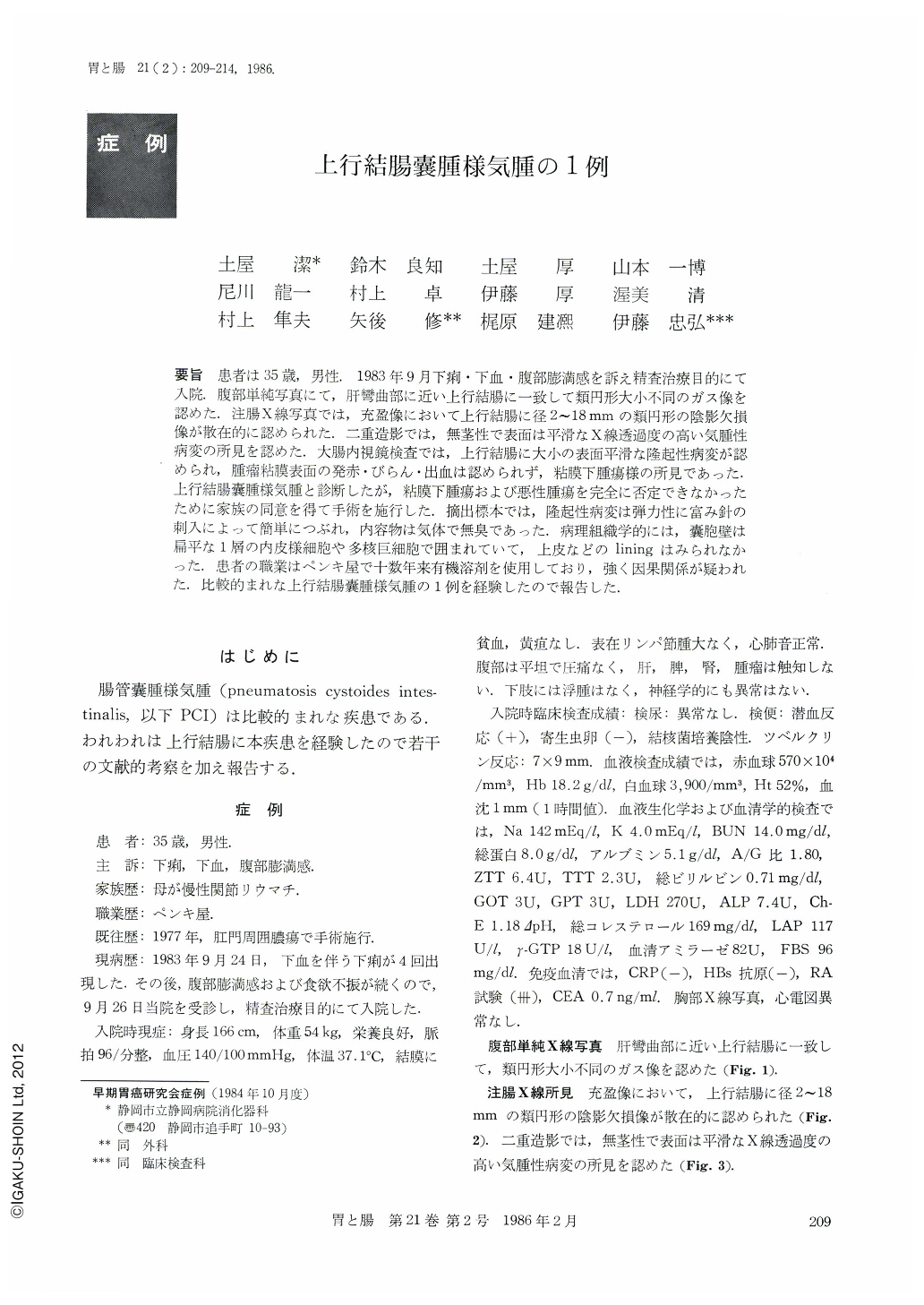
要旨 患者は35歳,男性.1983年9月下痢・下血・腹部膨満感を訴え精査治療目的にて入院.腹部単純写真にて,肝彎曲部に近い上行結腸に一致して類円形大小不同のガス像を認めた.注腸X線写真では,充盈像において上行結腸に径2~18mmの類円形の陰影欠損像が散在的に認められた.二重造影では,無茎性で表面は平滑なX線透過度の高い気腫性病変の所見を認めた.大腸内視鏡検査では,上行結腸に大小の表面平滑な隆起性病変が認められ,腫瘤粘膜表面の発赤・びらん・出血は認められず,粘膜下腫瘍様の所見であった.上行結腸囊腫様気腫と診断したが,粘膜下腫瘍および悪性腫瘍を完全に否定できなかったために家族の同意を得て手術を施行した.摘出標本では,隆起性病変は弾力性に富み針の刺入によって簡単につぶれ,内容物は気体で無臭であった.病理組織学的には,囊胞壁は扁平な1層の内皮様細胞や多核巨細胞で囲まれていて,上皮などのliningはみられなかった.患者の職業はペンキ屋で十数年来有機溶剤を使用しており,強く因果関係が疑われた.比較的まれな上行結腸囊腫様気腫の1例を経験したので報告した.
The patient, a 35-year-old man, came to our hospital for further minute check-up with complaints of diarrhea, melena and abdominal fullness in September 1983. Plain radiography of the abdomen showed almost round radiolucencies of varying size corresponding to the ascending colon near the hepatic flexure. Barium-filled roentgenogram of the colon showed almost round shadow defects, 2-18mm in diameter, scattered about in the ascending colon. Double contrast study of the colon showed a sessile pneumatic lesion with smooth surface, which was very radiolucent. Colonofiberscopic picture revealed elevated lesions of various size with smooth surface. On the surface of the lesions was seen no redness, erosion, or bleeding. The overall picture showed features of submucosal tumor. Although our diagnosis was pneumatosis cystoides of the ascending colon, we were unable to rule out submucosal tumor or some malignant tumor. With the consent of his family we decided to excise the lesions.
The resected specimens showed very elastic elevated lesions which were simply flattened with the insertion of a needle. The content was no other than air with no odor. Histologically, the wall of the cysts consisted of flattened endothelial cells and multinucleated cells with no lining such as epithelial cells.
The patient was a painter. The use of organic solvent for more than 10 years was strongly suspected for the cause of such lesions.

Copyright © 1986, Igaku-Shoin Ltd. All rights reserved.


